. 2004 Apr 27:4:5.
doi: 10.1186/1471-213X-4-5.

The roles of cis-inactivation by Notch ligands and of neuralized during eye and bristle patterning in Drosophila

Affiliations

The roles of cis-inactivation by Notch ligands and of neuralized during eye and bristle patterning in Drosophila

Yanxia Li et al. BMC Dev Biol. .

Abstract

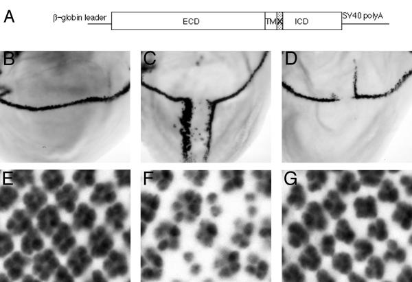
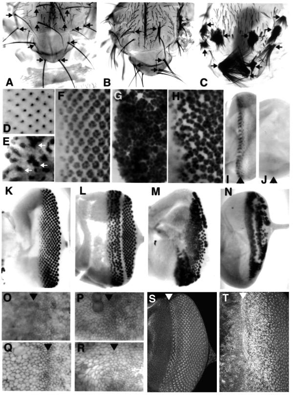
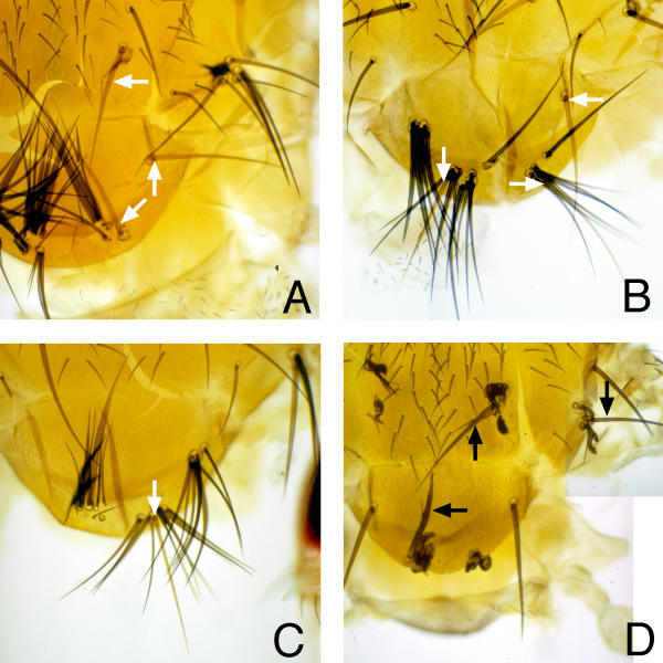
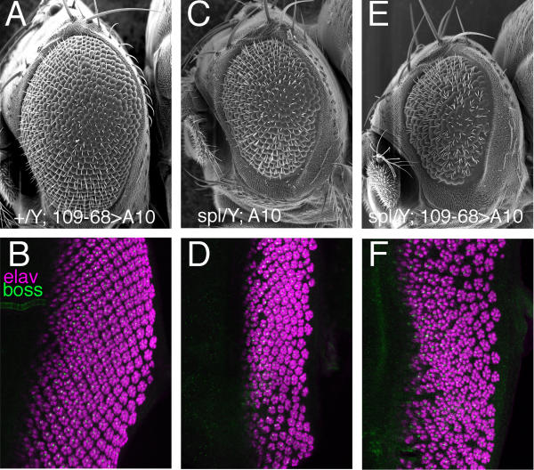
Background: The receptor protein Notch and its ligand Delta are expressed throughout proneural regions yet non-neural precursor cells are defined by Notch activity and neural precursor cells by Notch inactivity. Not even Delta overexpression activates Notch in neural precursor cells. It is possible that future neural cells are protected by cis-inactivation, in which ligands block activation of Notch within the same cell. The Delta-ubiquitin ligase Neuralized has been proposed to antagonize cis-inactivation, favoring Notch activation. Cis-inactivation and role of Neuralized have not yet been studied in tissues where neural precursor cells are resistant to nearby Delta, however, such as the R8 cells of the eye or the bristle precursor cells of the epidermis.

Results: Overexpressed ligands could block Notch signal transduction cell-autonomously in non-neural cells of the epidermis and retina, but did not activate Notch nonautonomously in neural cells. High ligand expression levels were required for cis-inactivation, and Serrate was more effective than Delta, although Delta is the ligand normally regulating neural specification. Differences between Serrate and Delta depended on the extracellular domains of the respective proteins. Neuralized was found to act cell nonautonomously in signal-sending cells during eye development, inconsistent with the view that Neuralized antagonizes cis-inactivation in non-neural cells.

Conclusions: Delta and Neuralized contribute cell nonautonomously to Notch signaling in neurogenesis, and the model that Neuralized antagonizes cis-inactivation to permit Notch activity and specification of non-neural cells is refuted. The molecular mechanism rendering Notch insensitive to paracrine activation in neural precursor cells remains uncertain.


Figures

Figure 1
Figure 1
Ectopic expression of Dl and Ser A. Outline of expression plasmids, incorporating β-globin leader sequences, SV40 poly A. An XbaI site at the cytoplasmic face of the transmembrane domain (X, shaded) allowed us to construct chimeric transgenes expressing the intracellular domain of one protein and extracellular and transmembrane domain of the other, as well as Dl and Ser transgenes with and without the XbaI site B-D. Cut protein induction at the developing dorso-ventral margin of wing imaginal discs dissected from white prepupae. B. wildtype. C. In dppGal4>SEDI ectopic wing margin is induced ventrally, as described previously for Ser. D. In dppGal4>DESI ectopic wing margin is induced dorsally, as described previously for Dl. E-G. Nuclear Elav protein in photoreceptor cells differentiating in the posterior eye imaginal disc. E. Wild type clusters contain 8 differentiating cells. F. In scaGal4>SESI the number of differentiating cells is reduced, as described previously for Ser. G. In scaGal4>DEDI the number of photoreceptors per cluster is also reduced.
Figure 2
Figure 2
Neurogenic effects A-C. Dorsal thorax preparations revealing pattern of bristles. A. Wildtype. The number and position of macrochaetae is highly stereotyped (arrows). B. Dl overexpression led to a few ectopic macrchaetae (arrows; see also Doherty et al., 1997). Shown is the strongest of our UAS-Dl insertion lines. Genotype is ScaG4>DESI (lineA10). C. Ser overexpression led to extreme neurogenic phenotypes apparently converting all cells in proneural regions to bristle precursors (arrows). Genotype is ScaG4>SESI. D-J Eye imaginal disc epithelia. D, E show eye discs labelled for the R8-specific protein Boss. Ser overexpression led to large clusters of R8-like cells (E) in place of an array of single cells in wildtype (D). F,G,H show eye discs with differentiating photoreceptor cells labelled for the neural-specific ELAV protein. F. In wildtype photoreceptors are progressively added until a total of 8 are differentiating in each cluster. G. Ser overexpression within and anterior to the morphogenetic furrow is strongly neurogenic, driving most cells posterior to the morphogenetic furrow into photoreceptor fate. H. Dl overexpression within and anterior to the morphogenetic furrow leads to a disorganized pattern of ommatidia (associated with the acceleration of the morphogenetic furrow) and is also weakly neurogenic. I,J Expression of the N target E(spl)mδ. I. wild type. J. Expression of E(spl)mδ, an important N target for lateral inhibition of R8, is lost when Ser is overexpressed in and anterior to the morphogenetic furrow. K-N. Eye discs labelled for ELAV expression reveals the extent of neural photoreceptor differentiation. All these discs were exposed to a 1 hour heat shock 22 hours before dissection. K. wild type. L. hs-Nintra. Photoreceptor differentiation is absent from a band of cells sensitive to N signaling at the time of the heat shock (arrow). M. Ser overexpression in and anterior to the morphogenetic furrow drives most cells posterior to the furrow into neural photoreceptor fates. N. Nintra expression is epistatic to ectopic Ser, preventing neural differentiation by a band of cells. O-R. Dl and N proteins on apical cell surfaces in the morphogenetic furrow region. Detergent-free antibody incubations only access protein on the cell surface. O. Wild type. Dl protein first appears on cell surfaces at the anterior of the morphogenetic furrow (arrow). P. Surface Dl protein is elevated and clearly detected both anterior and posterior to the furrow when expression is driven by the hH10transgene. Q. wild type. N protein outlines most eye disc cells. R. Similar levels of N protein reach the cell surface when Ser is overexpressed in and anterior to the furrow, even though these eye discs are strongly neurogenic due to reduced N function. S, T. Eye discs labelled for total Dl protein using detergent (bright fluorescent signal). S. wild type. An evolving pattern of Dl expression includes R8 precursors within groups of Dl-expressing cells just posterior to the morphogenetic furrow (arrowhead). T. hH10>SESI (same magnification; image recorded and processed identically to panel S). Dl protein reaches high levels in most eye disc cells. Note that Dl protein levels around the morphogenetic furrow (arrowhead) are higher than in any wild type cells.
Figure 3
Figure 3
Mosaic analysis of ligand-induced neurogenesis A-C. Mosaic thoraces from y hsFLP; FRT42 109-68 [y+]/FRT42; UAS-SESI flies. Yellow bristles (arrows) derive from recombinant cells lacking 109-68-dependent Ser expression. A maximum of one yellow bristle is seen in each proneural region. D. Mosaic thorax derived from f36a hsFLP; [abx>f+>Gal4]; UAS-DESI fly. Forked bristles derive from recombinant cells expressing the Dl transgene. Normal (not forked) bristles lack transgenic Dl expression (arrows). A maximum of one normal bristle is seen in each proneural region. These mosaics show that Ser and Dl overexpression are cell autonomously neurogenic and do not recruit neighboring cells to neural fates. They further suggest that each proneural region contains at least one cell not sensitive to high levels of ligand expression nearby.
Figure 4
Figure 4
Dl overexpression in R8 cells A. Overexpressing Dl in differentiating R8 cells leads to a rough eye of similar size to wild type (699 ± 21 ommatidia were recorded in male eyes and 790 ± 49 in females). Genotype is male 109-68>DESI (line A10). Overexpressing Dl in spl heterozygotes was similar (not shown; 693 ± 47 facets per eye were counted in spl/+; 109-68>A10 females, compared with 790 ± 49 in +/+;109-68>A10 females). 109-68 drives expression in R8 cells from column 1 onwards[54], too late to affect initial R8 specification but possibly in time to affect abnormal ommatidium development that occurs in spl mutants[7]. B. 109-68>DESI (line A10) eye discs labelled for the R8-specific Boss protein (green) and photoreceptor protein elav (magenta). No ommatidia or R8 cells were obviously missing as a result of Dl overexpression, nor were ectopic R8 cells seen. C. spl/Y; UAS DESI (line A10) males showed the small, rough eye typical of spl (386 ± 54 ommatidia). The Dl transgene is expected to remain unexpressed in this background. D. spl/Y; UAS DESI (line A10) eye discs show the typical spl phenotype. Some R8 cells and ommatidia are missing, other ommatidia have abnormal R8 cells and lack other photoreceptor cells[7]. E. spl/Y; 109-68>DESI (line A10) eyes. Dl overexpression in R8 cells did not rescue the spl mutant. In fact there was a statistically insignificant tendency for eyes tended to appear smaller than inspl. (312 ± 101 ommatidia were counted in spl/Y; 109-68> A10 male eyes). F. spl/Y; 109-68>DESI (line A10) eye discs also resembled spl. No rescue of the R8 or outer photoreceptor defects was apparent.
Figure 5
Figure 5
Mosaic analysis of neur A,B show phase-contrast micrographs of sections through eyes containing neur homozygous cells. The mutant cells lack pigment granules, which in photoreceptor cells are normally seen as dark bodies at the base of the rhabdomere. Photoreceptor cells are identified by number (R1, R2 etc) from salient ommatidia. Green numbers reflect morphologically normal, neur/+ or +/+ photoreceptors. Magenta indicates morphologically unaffected, neur mutant photoreceptors. Blue indicates morphologically transformed, neur/+ or +/+ photoreceptors. Panel A shoes several examples of neur mutant R3 cells that develop normally but are associated with R3>R4 transfromation by adjacent, neur/+ or +/+ cells. Panel B shows an ommatidium with neur mutant R1 and R6 cells. Although these cells appear unaffected, the neur/+ or +/+ cell in the R7 position adopted R1/6-like morphology.

Similar articles

Cited by

References

    1. Kimble J, Simpson P. The LIN-12/Notch signaling pathway and its regulation. Annu Rev Cell & Develop Biol. 1997;13:333–361. doi: 10.1146/annurev.cellbio.13.1.333. - DOI - PubMed
    1. Greenwald I. LIN-12/Notch signaling: lessons from worms and flies. Genes Dev. 1998;12:1751–1762. - PubMed
    1. Artavanis-Tsakonas S, Rand MD, Lake RJ. Notch signaling: cell fate control and signal integration in development. Science. 1999;284:770–776. doi: 10.1126/science.284.5415.770. - DOI - PubMed
    1. Kooh PJ, Fehon RG, Muskavitch MA. Implications of dynamic patterns of Delta and Notch expression for cellular interactions during Drosophila development. Development. 1993;117:493–507. - PubMed
    1. Baker NE. Notch signalling in the nervous system. Pieces still missing from the puzzle. BioEssays. 2000;22:264–273. doi: 10.1002/(SICI)1521-1878(200003)22:3<264::AID-BIES8>3.3.CO;2-D. - DOI - PubMed

Publication types

MeSH terms