Characterization of digital medical images utilizing support vector machines
- PMID: 15113418
- PMCID: PMC394338
- DOI: 10.1186/1472-6947-4-4
Characterization of digital medical images utilizing support vector machines
Abstract
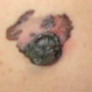
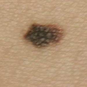
Background: In this paper we discuss an efficient methodology for the image analysis and characterization of digital images containing skin lesions using Support Vector Machines and present the results of a preliminary study.
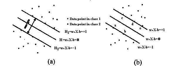
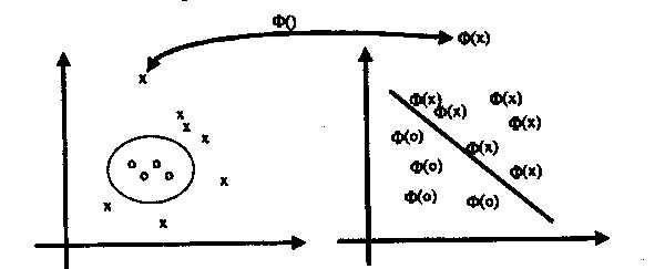
Methods: The methodology is based on the support vector machines algorithm for data classification and it has been applied to the problem of the recognition of malignant melanoma versus dysplastic naevus. Border and colour based features were extracted from digital images of skin lesions acquired under reproducible conditions, using basic image processing techniques. Two alternative classification methods, the statistical discriminant analysis and the application of neural networks were also applied to the same problem and the results are compared.
Results: The SVM (Support Vector Machines) algorithm performed quite well achieving 94.1% correct classification, which is better than the performance of the other two classification methodologies. The method of discriminant analysis classified correctly 88% of cases (71% of Malignant Melanoma and 100% of Dysplastic Naevi), while the neural networks performed approximately the same.
Conclusion: The use of a computer-based system, like the one described in this paper, is intended to avoid human subjectivity and to perform specific tasks according to a number of criteria. However the presence of an expert dermatologist is considered necessary for the overall visual assessment of the skin lesion and the final diagnosis.
Figures


Similar articles
-
Skin lesion classification using relative color features.Skin Res Technol. 2008 Feb;14(1):53-64. doi: 10.1111/j.1600-0846.2007.00261.x. Skin Res Technol. 2008. PMID: 18211602 Free PMC article.
-
An integrated computer supported acquisition, handling, and characterization system for pigmented skin lesions in dermatological images.IEEE Trans Inf Technol Biomed. 2005 Mar;9(1):86-98. doi: 10.1109/titb.2004.837859. IEEE Trans Inf Technol Biomed. 2005. PMID: 15787011 Clinical Trial.
-
Epiluminescence microscopy-based classification of pigmented skin lesions using computerized image analysis and an artificial neural network.Melanoma Res. 1998 Jun;8(3):261-6. doi: 10.1097/00008390-199806000-00009. Melanoma Res. 1998. PMID: 9664148
-
Distribution quantification on dermoscopy images for computer-assisted diagnosis of cutaneous melanomas.Med Biol Eng Comput. 2012 May;50(5):503-13. doi: 10.1007/s11517-012-0895-7. Epub 2012 Mar 22. Med Biol Eng Comput. 2012. PMID: 22438064 Review.
-
Dysplastic naevi: an update.Histopathology. 2010 Jan;56(1):112-20. doi: 10.1111/j.1365-2559.2009.03450.x. Histopathology. 2010. PMID: 20055909 Review.
Cited by
-
Automatic Detection of Malignant Melanoma using Macroscopic Images.J Med Signals Sens. 2014 Oct;4(4):281-90. J Med Signals Sens. 2014. PMID: 25426432 Free PMC article.
-
Classification of juvenile myoclonic epilepsy data acquired through scanning electromyography with machine learning algorithms.J Med Syst. 2012 Oct;36(5):2705-11. doi: 10.1007/s10916-011-9746-6. Epub 2011 Jun 17. J Med Syst. 2012. PMID: 21681512
-
Classification of parenchymal abnormality in scleroderma lung using a novel approach to denoise images collected via a multicenter study.Acad Radiol. 2008 Aug;15(8):1004-16. doi: 10.1016/j.acra.2008.03.011. Acad Radiol. 2008. PMID: 18620121 Free PMC article.
-
Computer-assisted diagnosis techniques (dermoscopy and spectroscopy-based) for diagnosing skin cancer in adults.Cochrane Database Syst Rev. 2018 Dec 4;12(12):CD013186. doi: 10.1002/14651858.CD013186. Cochrane Database Syst Rev. 2018. PMID: 30521691 Free PMC article.
-
HARLEY mitigates user bias and facilitates efficient quantification and co-localization analyses of foci in yeast fluorescence images.Sci Rep. 2022 Jul 18;12(1):12238. doi: 10.1038/s41598-022-16381-2. Sci Rep. 2022. PMID: 35851403 Free PMC article.
References
-
- Hall PN, Claridge E, Smith M. Computer screening for early detection of melanoma – is there a future?. Br J Dermatol. 1995;132:325–338. - PubMed
-
- Seidenari S, Burroni M, Dell'Eva G, Pepe P, Belletti B. Computerized evaluation of pigmented skin lesion images recorded by a videomicroscope: Comparison between polarizing mode observation and oil/slide mode observation. Skin Res Technol. 1995;1:187–191. - PubMed
MeSH terms
LinkOut - more resources
Full Text Sources