

p73alpha is a candidate effector in the p53 independent apoptosis pathway of cisplatin damaged primary murine colonocytes
- PMID: 15113856
- PMCID: PMC1770307
- DOI: 10.1136/jcp.2003.012559
p73alpha is a candidate effector in the p53 independent apoptosis pathway of cisplatin damaged primary murine colonocytes
Abstract
Aims: Colonocytes were derived from wild-type (wt) and p53 deficient mice to investigate p53 dependent and independent death pathways after cisplatin treatment, and the role of p53 in growth regulation of primary, untransformed epithelial cells.
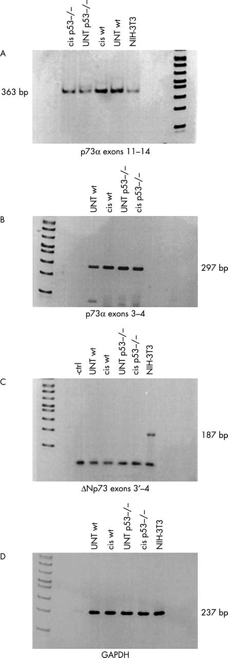
Methods: Wt and p53 null colonocytes were exposed to cisplatin and DNA synthesis, apoptosis, and p53, p21, and p73 expression were investigated after six, 12, and 24 hours. Major p73 isoforms were identified by reverse transcription polymerase chain reaction (RT-PCR).
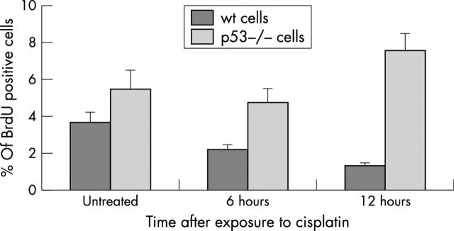
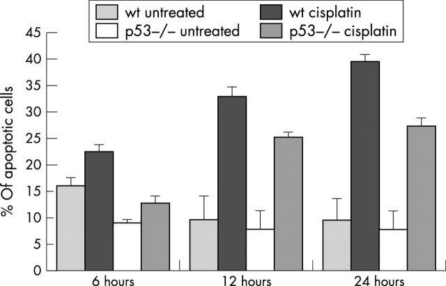
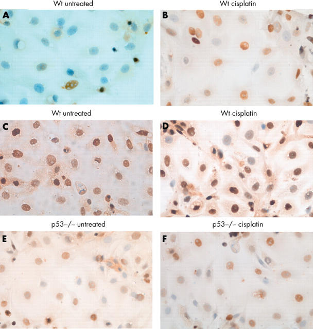
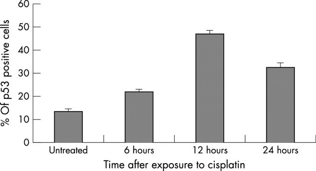
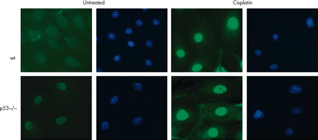
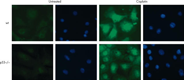
Results: Cisplatin treated wt cells exhibited cell cycle arrest, whereas p53 null cells continued to synthesise DNA, although both cell types died. Apoptosis was significantly higher in cisplatin treated wt and p53 null colonocytes than in controls at all timepoints, although apoptosis was lower in cisplatin treated p53 null colonocytes than in wt cells. p53 expression was upregulated in cisplatin treated wt colonocytes. p21 expression was high and remained unchanged in cisplatin treated wt cells, although it was reduced in the absence of p53. p73 was investigated because it could account for p53 independent p21 expression and p53 independent death. RT-PCR detected full length p73alpha. p73 transcript levels remained unchanged, whereas p73 protein accumulated in the nucleus of cisplatin treated cells, irrespective of genotype.
Conclusions: p53 is essential for cell cycle arrest, but not apoptosis in primary murine colonocytes. Apoptosis is reduced in cisplatin treated p53 null cells. Nuclear accumulation of endogenous p73 after cisplatin treatment suggests a proapoptotic role for p73alpha in the absence of p53 and collaboration with p53 in wt colonocytes.
Figures
Similar articles
-
Induction of p21 and nuclear accumulation of TAp73alpha and c-abl during apoptosis of cisplatin-treated primary pancreatic acinar cells.Int J Oncol. 2004 Dec;25(6):1661-70. Int J Oncol. 2004. PMID: 15547703
-
p73alpha regulates the sensitivity of bone marrow mesenchymal stem cells to DNA damage agents.Toxicology. 2010 Mar 30;270(1):49-56. doi: 10.1016/j.tox.2010.01.011. Epub 2010 Jan 25. Toxicology. 2010. PMID: 20100536
-
p73 function is inhibited by tumor-derived p53 mutants in mammalian cells.Mol Cell Biol. 1999 Feb;19(2):1438-49. doi: 10.1128/MCB.19.2.1438. Mol Cell Biol. 1999. PMID: 9891077 Free PMC article.
-
Choosing between growth arrest and apoptosis through the retinoblastoma tumour suppressor protein, Abl and p73.Biochem Soc Trans. 2001 Nov;29(Pt 6):666-73. doi: 10.1042/0300-5127:0290666. Biochem Soc Trans. 2001. PMID: 11709051 Review.
-
Role of p53 family members in apoptosis.J Cell Physiol. 2000 Feb;182(2):171-81. doi: 10.1002/(SICI)1097-4652(200002)182:2<171::AID-JCP5>3.0.CO;2-3. J Cell Physiol. 2000. PMID: 10623880 Review.
Cited by
-
Essential role of caspase-8 in p53/p73-dependent apoptosis induced by etoposide in head and neck carcinoma cells.Mol Cancer. 2011 Jul 31;10:95. doi: 10.1186/1476-4598-10-95. Mol Cancer. 2011. PMID: 21801448 Free PMC article.
-
Cisplatin-induced apoptosis in p53-deficient renal cells via the intrinsic mitochondrial pathway.Am J Physiol Renal Physiol. 2009 May;296(5):F983-93. doi: 10.1152/ajprenal.90579.2008. Epub 2009 Mar 11. Am J Physiol Renal Physiol. 2009. PMID: 19279129 Free PMC article.
-
Platinum Complexes in Colorectal Cancer and Other Solid Tumors.Cancers (Basel). 2021 Apr 25;13(9):2073. doi: 10.3390/cancers13092073. Cancers (Basel). 2021. PMID: 33922989 Free PMC article. Review.
References
-
- Iacopetta B. TP53 mutation in colorectal cancer. Hum Mutat 2003;21:271–6. - PubMed
-
- Kastan MB, Zhan Q, el-Deiry WS, et al. A mammalian cell cycle checkpoint pathway utilizing p53 and GADD45 is defective in ataxia-telangiectasia. Cell 1992;71:587–97. - PubMed
-
- Dameron KM, Volpert OV, Tainsky MA, et al. Control of angiogenesis in fibroblasts by p53 regulation of thrombospondin-1. Science 1994;265:1582–4. - PubMed
Publication types
MeSH terms
Substances
LinkOut - more resources
Full Text Sources
Molecular Biology Databases
Research Materials
Miscellaneous