. 2004 May;78(10):5045-55.
doi: 10.1128/jvi.78.10.5045-5055.2004.

Requirement for integrase during reverse transcription of human immunodeficiency virus type 1 and the effect of cysteine mutations of integrase on its interactions with reverse transcriptase

Affiliations

Requirement for integrase during reverse transcription of human immunodeficiency virus type 1 and the effect of cysteine mutations of integrase on its interactions with reverse transcriptase

Kai Zhu et al. J Virol. 2004 May.

Abstract

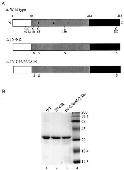
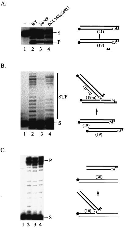
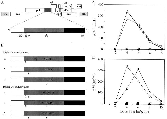
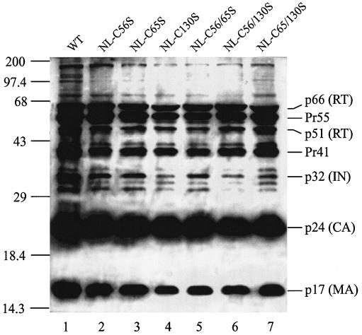
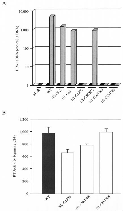
Retroviral integrase catalyzes the essential step of integrating a double-stranded DNA copy of the viral genome into a host cell chromosome. Mutational studies have revealed that integrase is involved in additional steps of viral replication, but the mechanism for the pleiotropic effect is not well characterized. Since Cys residues generally play crucial roles in protein structure and function, we introduced Cys-to-Ser substitutions at positions 56, 65, and 130 of human immunodeficiency virus type 1 (HIV-1) integrase to determine their effects on integration activity and viral replication. None of the substitutions significantly affected the enzymatic activities in vitro. When introduced into the NL4-3 molecular clone of HIV-1, mutant viruses encoding Cys mutations at positions 56 and 65 of integrase replicated similarly to the wild-type virus in CD4(+)-T-cell lines, whereas the C130S-containing virus was noninfectious. The entry and postintegration steps of the viral life cycle for all mutant viruses were normal, and all had particle-associated reverse transcriptase (RT) activity. However, early reverse-transcribed DNA products were absent in the lysate of cells infected with the C130S mutant virus, indicating that the mutation abolished the ability of the virus to initiate endogenous reverse transcription. Coimmunoprecipitation using purified integrase and RT showed that the C-terminal domain of wild-type HIV-1 integrase interacted with RT. The interaction between integrase and RT was not affected in the presence of a reducing or alkylating agent, suggesting that the interaction did not involve a disulfide linkage. The C130S substitution within the core region may disrupt the protein recognition interface of the C-terminal domain and abolish its ability to interact with RT. Our results indicate that integrase plays an important role during the reverse-transcription step of the viral life cycle, possibly through physical interactions with RT.


Figures

FIG. 1.
FIG. 1.
Recombinant wild-type and mutant HIV-1 INs. (A) Schematic representation of wild-type HIV-1 IN and Cys mutants used in the in vitro assays. N and C, N and C termini of the protein, respectively. The open boxes represent the N-terminal domain (amino acids 1 to 50) containing the conserved zinc-binding HHCC motif, the shaded boxes represent the central core domain (amino acids 51 to 212) containing the catalytic DD(35)E motif, and the solid boxes represent the C-terminal domain (amino acids 213 to 288). The six Cys residues and their amino acid positions are indicated in the wild-type IN (construct a). Replacements of Cys by Ser are indicated in both IN-NR (construct b) and IN-C56/65/280S (construct c). (B) Coomassie blue-stained SDS-polyacrylamide gel of 100 pmol of purified recombinant wild-type IN (WT; lane 1) and its mutant derivatives containing Cys-to-Ser substitutions at positions 56, 65, 130, and 280 (IN-NR; lane 2) or at positions 56, 65, and 280 (IN-C56/65/280S; lane 3). Molecular mass markers (Gibco) are shown in lane 4 and are labeled in kilodaltons on the right.
FIG. 2.
FIG. 2.
Enzymatic activities of wild-type and mutant HIV-1 INs in vitro. (A) 3′-end processing. (B) Strand transfer. (C) Disintegration. In all three panels, lane 1 contained the labeled substrate only, lane 2 had a reaction mixture containing the wild-type IN, and lanes 3 and 4 had reaction mixtures containing the mutant IN-NR and IN-C56/65/280S, respectively. The migration positions of the substrates (S) and products (P, 3′-end processing or disintegration product; STP, strand transfer product) are marked. For each in vitro assay, a model of the reaction substrate and products is at the right of the gel. The solid circles indicate the positions of the 32P label at the 5′ end of the oligonucleotide. The thick lines represent viral sequences, and the thin lines represent target sequences. The numbers in parentheses indicate the lengths in nucleotides of substrates and products.
FIG. 3.
FIG. 3.
Replication kinetics of mutant HIV-1 viral clones. (A) Genomic organization of HIV-1. The IN-coding sequence, represented by a dark shaded box, is expanded to show the positions of Cys residues. The IN domains are described in the legend to Fig. 1A. (B) HIV-1 clones containing a mutated IN. Viral clones encode either a single Cys-to-Ser mutation at amino acid residue position 56 (construct a), 65 (construct b), or 130 (construct c) or two Cys-to-Ser mutations at positions 56 and 65 (construct d), 56 and 130 (construct e), or 65 and 130 (construct f) of IN. (C) Replication kinetics of wild-type and single-Cys mutant viruses. CEM cells were infected with equal amounts of p24 equivalent of the wild type (○) or single-Cys mutant NL-C56S (Δ), NL-C65S (▿), or NL-C130S (□). (D) Replication kinetics of wild-type and double-Cys mutant viruses. CEM cells were infected with wild-type (○) or double-Cys mutant NL-C56/65S (▴), NL-C56/130S (▾), or NL-C65/130S (▪). In both panels C and D, the culture media were monitored for p24 production at the indicated time points postinfection. Mock infection was carried out with heat-inactivated wild-type virus (•).
FIG. 4.
FIG. 4.
Western blot analysis of HIV-1 proteins in viral particles. Each lane contains proteins from 50 ng of p24 equivalent of viral lysates separated on an SDS-12% polyacrylamide gel and probed with anti-HIV-1 human serum. Lane 1, wild-type virus (WT); lanes 2 to 7, various Cys mutant viruses as identified above the lanes. The numbers on the left are molecular masses in kilodaltons, and the labels on the right correspond to positions of viral proteins or their precursors.
FIG. 5.
FIG. 5.
cDNA synthesis and RT activities of wild-type and mutant viruses. (A) Quantitative analysis of viral cDNA synthesis by real-time PCR. The amount of early reverse-transcribed viral DNA present in CEM cells was measured 8 h after infection with the wild-type (WT) or mutant viruses (shaded bars). All viral stocks were treated with RNase-free DNase I to remove potential contamination by plasmid DNA. The culture medium alone was used in the mock infection (hatched bar), and equivalent amounts of the respective heat-inactivated viruses (solid bars) were used as negative controls for infection. For normalization of the DNA input of each sample, the human β-globin gene was amplified under identical PCR conditions. The results are expressed as the number of copies of HIV-1 cDNA detected per microgram of total DNA extracted from infected cells. The values shown represent the averages of two independent experiments. In each experiment, the standards and test samples were run in duplicate. (B) Particle-associated RT activities of wild-type and C130S-containing mutant viruses. After transient transfection of 293T cells, WT (shaded bar) and mutant (open bars) viruses were harvested, concentrated, lysed, and assayed for particle-associated RT activity as described in Materials and Methods. The result for each viral clone is shown as the mean ± standard error of three experiments using independently derived virus stocks.
FIG. 6.
FIG. 6.
Interaction between HIV-1 IN and RT. (A) Coimmunoprecipitation of RT and IN in vitro. Lane 1 contained the heterodimer purified RT (p66/p51), and IN and served as a positive control for immunoblotting and size markers. Lane 2 was a negative control and contained only the rabbit anti-HIV-1 RT polyclonal antibody. Lane 3 was a complete reaction without IN. For the coimmunoprecipitation experiments, wild-type HIV-1 IN (p32; lanes 8 and 9) or the Cys mutant derivative IN-NR (lanes 4 and 5) or IN-C56/65/280S (lanes 6 and 7) was incubated in the absence (lanes 4, 6, and 8) or presence (lanes 5, 7, and 9) of purified HIV-1 RT. The RT or RT-IN complex was immunoprecipitated with the rabbit anti-HIV-1 RT polyclonal antibody as described in Materials and Methods. The top blot was probed with the mouse anti-RT monoclonal antibody (αRT), and the bottom blot was probed with the rabbit anti-IN polyclonal antibody (αIN). The 50-kDa bands in lanes 2 to 9 correspond to the immunoglobulin G heavy chain (IgG-γ) that cross-reacted with the mouse anti-RT monoclonal antibody used for immunoblotting. (B) Mapping of the IN domain that interacts with RT. Various HIV-1 IN truncation mutants (as labeled above the lanes) were incubated with RT and immunoprecipitated using the rabbit anti-HIV-1 RT polyclonal antibody as described above. The blot was probed with anti-HIV-1 human serum.
FIG. 7.
FIG. 7.
RT-IN interaction does not involve disulfide bond formation. (A) Effect of a reducing agent on RT-IN interaction. HIV-1 IN was incubated in the absence (−) (lanes 1 and 3) or presence (+) (lanes 2 and 4) of RT and immunoprecipitated with a rabbit anti-HIV-1 RT polyclonal antibody. The immunoprecipitated complex was left untreated (lanes 1 and 2) or treated with β-ME (lanes 3 and 4), separated by SDS-PAGE, and analyzed by Western blotting using the rabbit anti-IN polyclonal antibody. The 64-kDa band in lane 2 corresponds to IN homodimers (5). The lower level of IN in lane 4 compared to lane 2 is due to a direct effect of β-ME on IN, possibly by altering the reactivity of IN to the anti-IN antibody. IgG-γ, immunoglobulin G heavy chain. (B) Effect of an alkylating agent on RT-IN interaction. Forty picomoles of HIV-1 IN in a final volume of 10 μl was preincubated at room temperature for 15 min in buffer C (20 mM HEPES-Na, pH 6.8, 250 mM NaCl, 0.2 mM MgCl2) with (lanes 1 and 2) or without (lanes 3 to 6) 10 mM DTT (20 mM equivalent of free sulfhydryls). The IN was then treated with 3.6 (2× molar excess; lanes 3 and 4) or 18 (10× molar excess; lanes 1, 2, 5, and 6) mM NEM for 1 h at 4°C. In lanes 3 to 6, the activity of NEM was quenched with 10 mM DTT. The samples were then added to buffer C with (lanes 2, 4, and 6) or without (lanes 1, 3, and 5) RT. Coimmunoprecipitation was carried out with the rabbit anti-HIV-1 RT polyclonal antibody. The immunoprecipitated complex was eluted with the buffer containing β-ME, and the rest of the procedure was as described for panel A. The extent of NEM modification was monitored using electrospray ionization-mass spectrometry. At 2× molar excess of NEM, all INs were modified, and the distribution of INs containing four, five, and six NEM adducts was ∼35, 50, and 10%, respectively (data not shown).The IN with four NEM adducts likely represented Cys modification at positions 56, 65, 130, and 280, since the Cys residues at positions 40 and 43 are bound with zinc and therefore should be much less reactive. The extent of NEM adduct formation at 10× excess was not determined.

Similar articles

Cited by

References

    1. Adachi, A., H. E. Gendelman, S. Koenig, T. Folks, R. Willey, A. Rabson, and M. A. Martin. 1986. Production of acquired immunodeficiency syndrome-associated retrovirus in human and nonhuman cells transfected with an infectious molecular clone. J. Virol. 59:284-291. - PMC - PubMed
    1. Aiken, C., and D. Trono. 1995. Nef stimulates human immunodeficiency virus type 1 proviral DNA synthesis. J. Virol. 69:5048-5056. - PMC - PubMed
    1. Aldovini, A., and B. D. Walker. 1990. Techniques in HIV research. Stockton Press, New York, N.Y.
    1. Ausubel, F. A., R. Brent, R. E. Kingston, D. D. Moore, J. G. Seidman, J. A. Smith, and K. Struhl (ed.). 1999. Current protocols in molecular biology. John Wiley & Sons, Inc., New York, N.Y.
    1. Bischerour, J., H. Leh, E. Deprez, J. C. Brochon, and J. F. Mouscadet. 2003. Disulfide-linked integrase oligomers involving C280 residues are formed in vitro and in vivo but are not essential for human immunodeficiency virus replication. J. Virol. 77:135-141. - PMC - PubMed

Publication types