. 2004 May;78(10):5056-67.
doi: 10.1128/jvi.78.10.5056-5067.2004.

Interaction between human immunodeficiency virus type 1 reverse transcriptase and integrase proteins

Affiliations

Interaction between human immunodeficiency virus type 1 reverse transcriptase and integrase proteins

Eric A Hehl et al. J Virol. 2004 May.

Abstract

Reverse transcriptase (RT) and integrase (IN) are two key catalytic enzymes encoded by all retroviruses. It has been shown that a specific interaction occurs between the human immunodeficiency virus type 1 (HIV-1) RT and IN proteins (X. Wu, H. Liu, H. Xiao, J. A. Conway, E. Hehl, G. V. Kalpana, V. R. Prasad, and J. C. Kappes, J. Virol. 73:2126-2135, 1999). We have now further examined this interaction to map the binding domains and to determine the effects of interaction on enzyme function. Using recombinant purified proteins, we have found that both a HIV-1 RT heterodimer (p66/p51) and its individual subunits, p51 and p66, are able to bind to HIV-1 IN. An oligomerization-defective mutant of IN, V260E, retained the ability to bind to RT, showing that IN oligomerization may not be required for interaction. Furthermore, we report that the C-terminal domain of IN, but not the N-terminal zinc-binding domain or the catalytic core domain, was able to bind to heterodimeric RT. Deletion analysis to map the IN-binding domain on RT revealed two separate IN-interacting domains: the fingers-palm domain and the carboxy-terminal half of the connection subdomain. The carboxy-terminal domain of IN alone retained its interaction with both the fingers-palm and the connection-RNase H fragments of RT, but not with the half connection-RNase H fragment. This interaction was not bridged by nucleic acids, as shown by micrococcal nuclease treatment of the proteins prior to the binding reaction. The influences of IN and RT on each other's activities were investigated by performing RT processivity and IN-mediated 3' processing and joining reactions in the presence of both proteins. Our results suggest that, while IN had no influence on RT processivity, RT stimulated the IN-mediated strand transfer reaction in a dose-dependent manner up to 155-fold. Thus, a functional interaction between these two viral enzymes may occur during viral replication.


Figures

FIG. 1.
FIG. 1.
Native IN interacts with all three forms of HIV-1 RT. Hexahistidine-tagged RT heterodimer (p66/p51), p51, and p66 bound to Ni2+-NTA agarose beads were incubated with crude bacterial lysates containing native IN. The beads were washed, and the bound proteins were resolved by SDS-15% PAGE. (Top) Immunoblot probed with polyclonal α-IN antibodies directed to N-terminal residues 23 to 34. Lanes RT, IN, and ΔIN contain unreacted lysates from bacteria (RT), the IN expression plasmid (pT7IN), and the expression plasmid without the IN sequences (pT7ΔIN). Lanes 1 and 2, empty Ni2+-NTA beads incubated with pT7ΔIN lysate or pT7IN lysate; lanes 3 to 5, Ni2+-NTA beads bound to p66/p51, p51, or p66; lanes 6 to 8, Ni2+-NTA beads bound to p66/p51, p51, or p66 incubated with pT7ΔIN lysate; lanes 9 to 11, Ni2+-NTA beads bound to p66/p51, p51, or p66 incubated with pT7IN lysate. (Bottom) Parallel protein transfer blot probed with the α-RT antibody 8C4D7 to ensure the presence of similar input RT bait protein. Lanes 1 to 11 are identical to those in panel A.
FIG. 2.
FIG. 2.
Multimerization of IN is not critical to RT-IN interaction. (A) Lanes 1 to 3, empty, GST-bound, and GST-IN-bound G beads incubated with bacterial lysates containing RT p66; lane 4, GST-IN/V260E-bound G beads incubated with p66 RT lysate. The immunoblot was probed with the α-RT antibody 5B2B2. (B) Parallel SDS-PAGE gel from the experiment shown in panel A Coomassie stained to ensure the presence of equivalent amounts of the proteins.
FIG. 3.
FIG. 3.
Mapping the IN-binding domain on HIV-1 RT. In experiments similar to those shown in Fig. 2, G beads bound to GST or GST-IN were incubated with bacterial lysates containing various truncation mutants of RT and washed, and the bound proteins were resolved on duplicate SDS-PAGE gels. The proteins on one gel were transferred to nitrocellulose and subjected to immunostaining with α-RT antibodies, and the second gel was stained with Coomassie blue to ensure equal input of the bait proteins. (A) Pull-down experiments to map the IN-binding domains on RT p66. Lanes 1, 5, 7, 9, 11, and 13 contained bound proteins from a control incubation of GST bound to G beads, and lanes 2 to 4, 6, 8, 10, 12, and 14 to 17 contained proteins bound to GST-IN bound to G beads. RT mutants were added to the lanes as follows: lane 2, p66; lane 3, TCR; lane 4, Conn-R; lanes 5 and 6, C*R; lanes 7 and 8, R; lanes 9 and 10, p66; lanes 11 and 12, T; lanes 13 and 14, p66; lane 15, p51; lane 16, FPT; and lane 17, FP. The corresponding lanes on the bottom show stained protein indicating the input bait protein levels. The differences in p66 intensities in lanes 2 and 14 are due to different antibodies used for the Western blots. (B) A schematic summarizing results obtained in the experiment shown in panel A. The horizontal bar at the top represents full-length RT p66, with various subdomains and their boundaries indicated by amino acid residue numbers. The RT truncations used in the pull-down experiment, their boundaries, and their abilities to bind IN are indicated. Below, the proposed domains of IN interaction are indicated. +, able to bind; −, not able to bind.
FIG. 4.
FIG. 4.
Mapping the RT-binding domain on IN. (A) In an experimental setup for pull-down experiments similar to that in Fig. 3, GST-IN and GST fusions of various truncations of IN were incubated with heterodimeric RT, followed by washing and resolving the bound proteins on SDS-PAGE. As before, one gel was transferred to nitrocellulose and probed with monoclonal α-RT antibody 5B2B2 (top), and a duplicate gel with the same proteins was stained with Coomassie blue (bottom). Lane 1, empty G beads; lane 2: GST-bound G beads; lanes 3 and 7, GST-IN-bound G beads; lanes 4, 5, 6, and 8, G beads bound to GST-IN Zn finger domain (amino acid residues 1 to 50), GST-IN catalytic core domain (residues 48 to 208), GST-IN C-terminal domain (residues 201 to 288), and GST-IN C-terminal deletion (residues 1 to 220), respectively. (B) Schematic showing a summary of RT-binding abilities of full-length IN and the various truncation mutants of IN tested. The amino acid residues of each mutant are shown at the left of the horizontal bar representing each deletion. +, able to bind; −, not able to bind.
FIG. 5.
FIG. 5.
Interaction between the binding domains occurs without nucleic acid bridging. G beads, GST, or GST-IN prepared with or without micrococcal-nuclease treatment were incubated with p66, FP, Conn-R, or C*R similarly prepared with or without micrococcal-nuclease treatment. (A) Pull-down reactions using micrococcal-nuclease-treated interaction partners. Plain G beads (lane 1) or G beads bound to 1 μg of GST (lane 2) or GST-IN (lane 3) were incubated with p66 protein. G beads bound to GST-IN alone were incubated with the FP domain (lane 4). Subsequent to the pull-down and SDS-PAGE (15% acrylamide) analysis of the bound proteins, an immunoblot was prepared and probed with the monoclonal antibody 8C4D7, which recognizes the epitope located between RT residues 193 and 284, which overlaps with the FP domains. (B) Pull-down reactions using micrococcal-nuclease-treated interaction partners. Similar to panel A, plain G beads (lane 5) or G beads bound to GST (lane 6) or GST-IN (lane 7) were incubated with p66. The G beads bound to GST-IN were also separately incubated with Conn-R (lane 8) or C*R (lane 9). Although both panels A and B are from the same SDS-PAGE gel, the immunoblot corresponding to panel B was probed with a different monoclonal antibody directed to the R domain (residues 440 to 560 of RT p66) to facilitate detection of the C-terminal fragments of RT. (C and D) The lanes are identical to those in panels A and B, except that none of the proteins were treated with micrococcal nuclease. (E and F) The lanes are similar to those in panels A and B, except that GST-C-terminal IN was used instead of GST-IN. All proteins were also treated with micrococcal nuclease. (G and H) The lanes are similar to those in panels E and F in that GST-C-terminal IN was used for pull downs, but no proteins were treated with micrococcal nuclease. The numbers on the left of the panels are molecular weight markers (prestained Invitrogen Benchmark).
FIG. 6.
FIG. 6.
W235 substitutions do not disrupt RT-IN interaction. MBP-IN fusion proteins containing W235A or W235E mutations were concentrated from induced lysates by using amylose resin beads. Amylose resins alone (lanes 1) and amylose bound to MBP (lanes 2), MBP-INW235A (lanes 3), or MBP-INW235E (lanes 4) protein were incubated with lysates containing HIV-1 RT heterodimer followed by washing the resin and analysis of bound proteins on SDS-PAGE. (A) Immunoblot analysis using α-RT antibodies. The positions of 66- and 51-kDa subunits of RT are indicated. (B) Gel identical to that in panel A stained with Coomassie brilliant blue. The migration positions of the MBP-IN fusion and MBP are indicated.
FIG. 7.
FIG. 7.
Effects of mutations at conserved IN residues on RT-IN interaction. GST pull-down experiments were carried out as before by incubating wild-type GST-IN or the substitution mutants H12A, H16A, D116A, and F185A with lysates containing heterodimeric RT. (A) Bound proteins were resolved on SDS-PAGE, transferred to nitrocellulose, and probed with the α-RT antibody 5B2B2. Lanes 1 to 3, empty, GST-bound, and GST-IN-bound G beads; lanes 4 to 7, GST-IN-H12A, GST-IN-H16A, GST-IN-D116A, and GST-IN-F185A, respectively. The positions of p66 and p51 polypeptides are indicated. (B) A parallel SDS-PAGE gel was Coomassie blue stained to ensure the presence of equivalent inputs of proteins. The positions of GST and GST-IN are indicated.
FIG. 8.
FIG. 8.
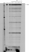
Effect of IN on the processivity of HIV-1 RT. Using a primer that binds to the primer binding site, processivity reactions were done in the presence or absence of IN. Lane 1, primer alone; lane 2, untrapped reaction in the absence of IN; lane 3, pretrapped reaction where RT was mixed with poly(rA) · oligo(dT) prior to the addition of template-primer to ensure the effectiveness of the trap; lane 4, reaction in which dNTPs and trap were added simultaneously; lanes 5 to 12, reactions in which dNTPs and trap were added simultaneously in the presence of increasing amounts of IN ranging from a 1:0.03125 to a 1:4 molar ratio of RT to IN (with a constant input of RT). nt, nucleotides.
FIG. 9.
FIG. 9.
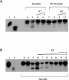
Effect of RT on 3′ processing by IN protein. (A) Using radiolabeled U5.3/U5.4 substrate, a comparison was made between normal IN buffer and RT-IN buffer. Lane 1, unclipped U5.3/U5.4; lane 2, U5.5/U5.4, the preclipped version of U5.3/U5.4; lane 3, 3′ processing reaction using IN buffer in the absence of IN; lane 4, processing reaction using IN buffer in the presence of IN; lanes 5 and 6, processing reactions using IN buffer in the presence of IN and RT in a 1:1 to 1:4 molar ratio of IN monomer to RT, respectively; lanes 7 to 10, exactly as for lanes 3 to 6 except using RT-IN buffer. The reactions in lanes 4 to 6 and 8 to 10 were performed with a constant input of IN. (B) All of the 3′ processing reactions were done using normal IN buffer. Lanes 1 and 2, same as for panel A; lane 3, 3′ processing reaction in the absence of IN; lane 4, 3′ processing reaction in the absence of IN and the presence of 9.36 pmol of RT; lane 5, 3′ processing reaction in the presence of 4.68 pmol of IN monomer; lanes 6 to 12, 3′ processing reactions in the presence of 4.68 pmol of IN monomer and the following concentrations of RT: 1:0.03125, 1:0.0625, 1:0.125, 1:0.25, 1:0.5, 1:1, and 1:2 (IN monomer to RT heterodimer), respectively.
FIG. 10.
FIG. 10.
Effect of RT on the joining reaction by IN. (A) Effect of RT on joining reactions by IN using standard IN buffer. (Top) Autoradiogram displaying products of joining reactions carried out in the presence of increasing concentrations of RT. Lane 1, no IN; lane 2, RT alone (4.68 pmol); lane 3, IN alone (4.68 pmol); lanes 4 to 7, 4.68 pmol of IN with various proportions of RT at molar ratios (IN monomer to RT heterodimer) of 1:0.125, 1:0.25, 1:0.5, and 1:1, respectively. (Bottom) Agarose gel of the same joining reactions. Lane M, markers showing 4-, 3-, 2-, and 1.5-kb bands; lane 0, 0.2 μg of uncut pBluescript. Lanes 1 to 7 are identical to lanes 1 to 7 above. The starting supercoiled DNA substrate (bottom only; indicated with a twisted circle symbol) and the nonconcerted joining reaction products (top and bottom; indicated with a panhandle symbol) are shown. (B) (Top and bottom) Exactly as described for panel A, except that the reactions were carried out in the presence of RT-IN buffer and the marker lane (M) shows only 3-, 2-, and 1.5-kb bands.

Similar articles

Cited by

References

    1. Andrake, M. D., and A. M. Skalka. 1995. Multimerization determinants reside in both catalytic core and C terminus of avian sarcoma virus integrase. J. Biol. Chem. 270:29299-29306. - PubMed
    1. Arts, E. J., X. Li, Z. Gu, L. Kleiman, M. A. Parniak, and M. A. Wainberg. 1994. Comparison of deoxyoligonucleotide and tRNA(Lys-3) as primers in an endogenous human immunodeficiency virus-1 in vitro reverse transcription/template-switching reaction. J. Biol. Chem. 269:14672-14680. - PubMed
    1. Asante-Appiah, E., and A. M. Skalka. 1999. HIV-1 integrase: structural organization, conformational changes, and catalysis. Adv. Virus Res. 52:351-369. - PubMed
    1. Beard, W. A., and S. H. Wilson. 1993. Kinetic analysis of template-primer interactions with recombinant forms of HIV-1 reverse transcriptase. Biochemistry 32:9745-9753. - PubMed
    1. Bukrinsky, M. I., N. Sharova, T. L. McDonald, T. Pushkarskaya, W. G. Tarpley, and M. Stevenson. 1993. Association of integrase, matrix, and reverse transcriptase antigens of human immunodeficiency virus type 1 with viral nucleic acids following acute infection. Proc. Natl. Acad. Sci. USA 90:6125-6129. - PMC - PubMed

Publication types

LinkOut - more resources