. 2004 May;78(10):5157-69.
doi: 10.1128/jvi.78.10.5157-5169.2004.

Effect of extension of the cytoplasmic domain of human immunodeficiency type 1 virus transmembrane protein gp41 on virus replication

Affiliations

Effect of extension of the cytoplasmic domain of human immunodeficiency type 1 virus transmembrane protein gp41 on virus replication

Woan-Eng Chan et al. J Virol. 2004 May.

Abstract

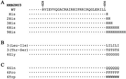
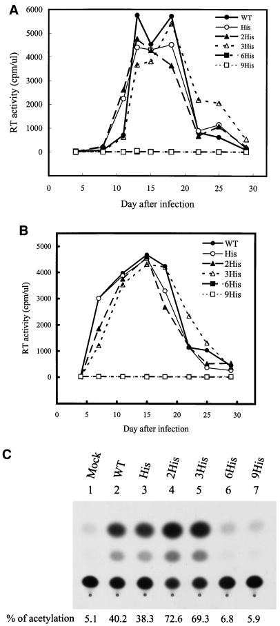
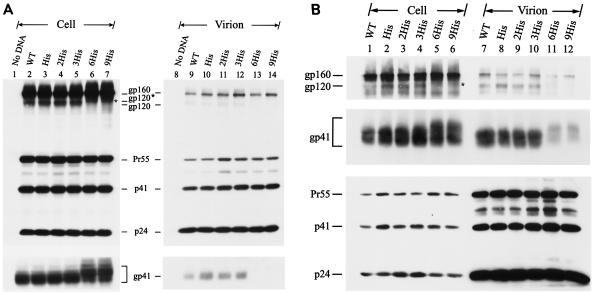
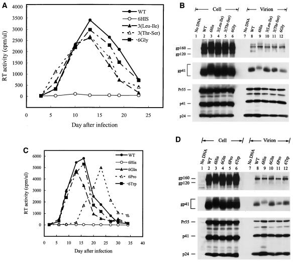
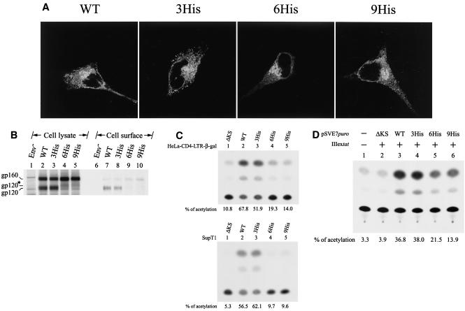
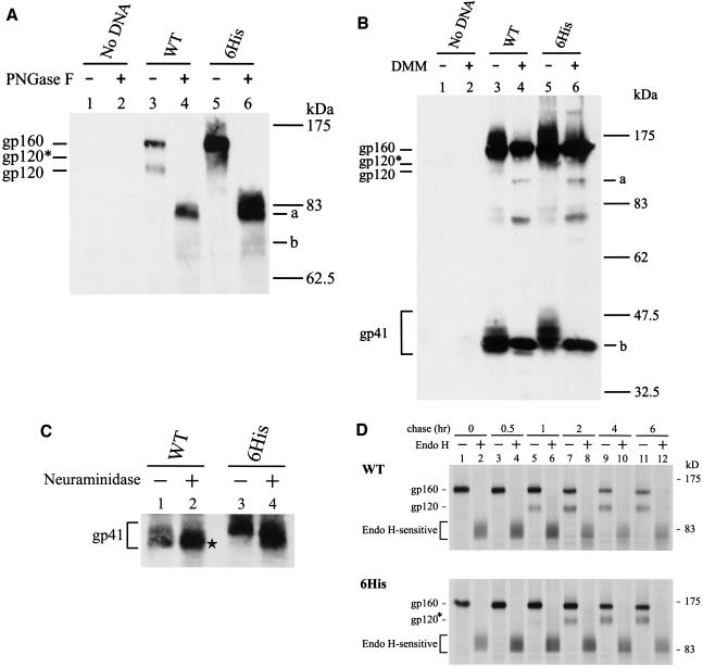
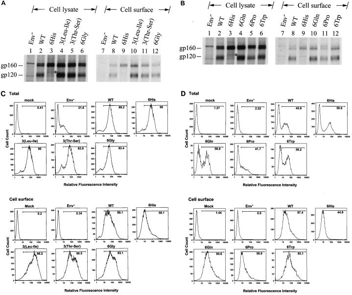
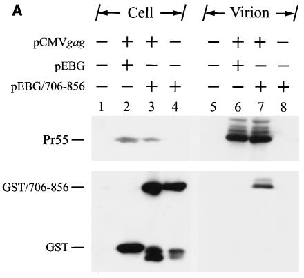
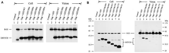
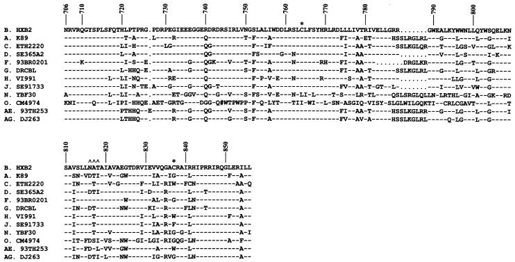
The biological significance of the presence of a long cytoplasmic domain in the envelope (Env) transmembrane protein gp41 of human immunodeficiency virus type 1 (HIV-1) is still not fully understood. Here we examined the effects of cytoplasmic tail elongation on virus replication and characterized the role of the C-terminal cytoplasmic tail in interactions with the Gag protein. Extensions with six and nine His residues but not with fewer than six His residues were found to severely inhibit virus replication through decreased Env electrophoretic mobility and reduced Env incorporation compared to the wild-type virus. These two mutants also exhibited distinct N glycosylation and reduced cell surface expression. An extension of six other residues had no deleterious effect on infectivity, even though some mutants showed reduced Env incorporation into the virus and/or decreased cell surface expression. We further show that these elongated cytoplasmic tails in a format of the glutathione S-transferase fusion protein still interacted effectively with the Gag protein. In addition, the immediate C terminus of the cytoplasmic tail was not directly involved in interactions with Gag, but the region containing the last 13 to 43 residues from the C terminus was critical for Env-Gag interactions. Taken together, our results demonstrate that HIV-1 Env can tolerate extension at its C terminus to a certain degree without loss of virus infectivity and Env-Gag interactions. However, extended elongation in the cytoplasmic tail may impair virus infectivity, Env cell surface expression, and Env incorporation into the virus.


Figures

FIG. 1.
FIG. 1.
Construction of HIV-1 cytoplasmic tail elongation mutants. The amino acid sequence in the single-letter code from residues 828 to 856 of Env of strain HXB2 is shown at the top. Extensions with different numbers of His residues (A) and residues other than His (B and C) at the C terminus of this domain were constructed as described in Materials and Methods. WT, wild type.
FIG. 2.
FIG. 2.
Replication kinetics of His mutants in CD4+ T cells. Cell-free wild-type (WT) and mutant viruses were used to challenge 106 CEM-SS cells (A) or SupT1 cells (B). Virus production as measured by reverse transcriptase (RT) activity was monitored after infection. Wild-type and mutant viruses were also used to infect 106 H938 cells, and CAT activity was monitored (C).
FIG. 3.
FIG. 3.
Viral protein expression of His mutants. (A) HeLa cells were transfected with the wild-type (WT) or mutant proviruses, and equal volumes of cell and virion lysates were analyzed by Western blotting with MAbs 183 and 902 (top panel). After stripping off of the membrane-bound antibodies, the same blot was reprobed with MAb Chessie 8 (bottom panel). (B) SupT1 cells were infected by wild-type and His mutant VSV G pseudotypes, and cell and virion lysates were analyzed by Western blotting with MAbs 902, Chessie 8, and 183, respectively. In each figure, the intracellular gp120 band observed in the 6His and 9His mutants is marked by an asterisk.
FIG. 4.
FIG. 4.
Replication and expression of cytoplasmic tail elongation mutants. (A and B) Analyses of the 3(Leu-Ile), 3(Thr-Ser), and 6Gly mutants. Cell-free wild-type (WT) and mutant viruses were used to challenge CEM-SS cells, and reverse transcriptase (RT) activity was monitored after infection (A). HeLa cells were transfected with the wild-type and mutant proviruses, and cell and virion lysates were analyzed by Western blotting with MAbs 902, Chessie 8, and 183 (B). (C and D) Analyses of the 6Gln, 6Pro, and 6Trp mutants. Replication of wild-type and mutant viruses in CEM-SS cells was assessed as described for panel A (C). Viral protein expression in HeLa cells was examined as described for panel B (D).
FIG. 5.
FIG. 5.
Analyses of His mutants. (A) Cellular localization of His mutants. HeLa cells cotransfected with pIIIextat and the wild-type (WT) and His mutant pSVE7puro plasmids were analyzed by confocal microscopy as described in Materials and Methods. (B) Cell surface biotinylation of the 6His mutant. HeLa cells transfected with pHXBCATΔBgl (3), marked Env, which do not express a functional env gene, and wild-type or mutant proviruses as indicated were metabolically labeled and surface biotinylated, and cell lysates were precipitated with anti-HIV-1 preadsorbed to protein A-Sepharose. Equal amounts of the isolated Env proteins were subjected to SDS-PAGE (lanes 1 to 5) or precipitated with neutravidin-agarose prior to SDS-PAGE (lanes 6 to 10). (C) env trans-complementation assay of the His mutants. Cell-free, env-defective HIV-1 cat reporter viruses generated in the presence of wild-type or mutant Env coexpression were used to challenge HeLa-CD4-LTR-β-gal (top panel) and SupT1 (bottom panel) cells, and CAT activity was assessed. (D) Quantitation of the syncytium-forming ability of the mutant proteins. 293T cells were cotransfected with pIIIextat and the ΔKS, wild-type, and mutant pSVE7puro plasmids. One million H938 cells were added to transfected cells 1 day after transfection, and cell lysates were assayed for CAT activity after 2 days of coculture.
FIG. 6.
FIG. 6.
Analyses of N-linked glycosylation of the 6His mutant. (A) PNGase F analysis. Equal volumes of MAb 902-isolated Env proteins from HeLa cells transfected with the wild-type (WT) or 6His mutant provirus were treated or not with PNGase F prior to Western blotting with MAb 902. Bands a and b represent the gp160 and gp120 forms without N-glycans, respectively. (B) DMM treatment. Cells transfected with the wild-type or mutant provirus were grown in the presence or absence of 1 mM DMM for 48 h, and cell lysates were analyzed by Western blotting with MAb 902. Bands a and b represent the high-mannose forms of gp120 and gp41, respectively. (C) Neuraminidase digestion. Equal volumes of the isolated wild-type and 6His mutant Env proteins were treated or not with neuraminidase prior to Western blotting with MAb Chessie 8. (D) Pulse-chase and endo H digestion. HeLa cells transfected with the wild-type or 6His mutant provirus were metabolically labeled and chased for different times. Equal volumes of MAb 902-isolated Env proteins were treated or not with endo H prior to Western blotting with MAb 902.
FIG. 7.
FIG. 7.
Cell surface expression of mutants containing six extra residues at the C terminus of the cytoplasmic tail. (A and B) Surface biotinylation of the 6His, 3(Leu-Ile), 3(Thr-Ser), and 6Gly mutants and of the 6His, 6Gln, 6Pro, and 6Trp mutants, respectively. HeLa cells transfected with the wild-type (WT) or mutant proviruses as indicated were metabolically labeled and surface biotinylated. Equal volumes of anti-HIV-1-isolated Env proteins were resolved by SDS-PAGE without (lanes 1 to 6) or with (lanes 7 to 12) prior precipitation with neutravidin-agarose. (C and D) FACS analyses of the 6His, 3(Leu-Ile), 3(Thr-Ser), and 6Gly mutants and of the 6His, 6Gln, 6Pro, and 6Trp mutants, respectively. VSV G-pseudotyped wild-type and mutant viruses as indicated were used to challenge CEM-SS cells, and total and cell surface Env expression was analyzed by FACS. Mock infection and infection with pseudotyped virus produced from pHXBCATΔBgl cotransfection, labeled Env, were used as negative controls.
FIG. 8.
FIG. 8.
Examination of Gag-cytoplasmic tail interactions. (A) Incorporation of a cytoplasmic tail fusion protein into Gag particles. 293T cells were cotransfected with pCMVgag together with pEBG or pEBG/706-856 as indicated. Cell and virion lysates were analyzed by Western blotting with MAb 183 and a rabbit anti-GST antibody, respectively. Transfection with pEBG/706-856 in the presence of pCDNA3 was also performed in parallel (lanes 4 and 8). (B) Isolation of the immature HIV-1 core structure from Gag and GST/706-856 coexpression. Cell-free, concentrated viral particles obtained from Gag and GST/706-856 coexpression were sedimented through a linear 30 to 70% sucrose gradient containing a layer of 1% Triton X-100. Isolated core particles in each fraction were analyzed by Western blotting with MAbs 183 and Chessie 8. (C). Sedimentation analysis of GST/706-856. 293T cells expressing GST/706-856 were lysed with phosphate-buffered saline containing 1% Triton X-100, and the lysates were sedimented through a sucrose gradient lacking detergent. After fractionation, proteins in each fraction was concentrated by 10% cold trichloroacetic acid precipitation and then analyzed by Western blotting with Chessie 8. (D) Lack of GST/706-856 incorporation into the 30LE mutant virus-like Gag particles. Cells were cotransfected with wild-type or 30LE mutant pHIVgptGag in the presence of pEBG or pEBG/706-856. Cell and virus lysates were analyzed with MAb 183 and rabbit anti-GST, respectively, by Western blotting. (E) Lack of interaction of GST/706-856 with the 30LE mutant Gag. 293T cells were cotransfected with pHIVgptΔGag or with wild-type or 30LE mutant pHIVgptGag in the presence of pEBG or pEBG/706-856. Transfected cells were lysed with Tris-buffered saline containing 10 mM 3-[(cholamidopropyl)-dimethylammonio]-1-propanesulfonate (CHAPS). One fraction of the cell lysates was subjected directly to Western blotting with rabbit anti-GST and MAb 183 (top panels). Another fraction was first incubated with glutathione-Sepharose 4B, and the bound proteins were analyzed by Western blotting with anti-GST and MAb 183 (bottom panel).
FIG. 8.
FIG. 8.
Examination of Gag-cytoplasmic tail interactions. (A) Incorporation of a cytoplasmic tail fusion protein into Gag particles. 293T cells were cotransfected with pCMVgag together with pEBG or pEBG/706-856 as indicated. Cell and virion lysates were analyzed by Western blotting with MAb 183 and a rabbit anti-GST antibody, respectively. Transfection with pEBG/706-856 in the presence of pCDNA3 was also performed in parallel (lanes 4 and 8). (B) Isolation of the immature HIV-1 core structure from Gag and GST/706-856 coexpression. Cell-free, concentrated viral particles obtained from Gag and GST/706-856 coexpression were sedimented through a linear 30 to 70% sucrose gradient containing a layer of 1% Triton X-100. Isolated core particles in each fraction were analyzed by Western blotting with MAbs 183 and Chessie 8. (C). Sedimentation analysis of GST/706-856. 293T cells expressing GST/706-856 were lysed with phosphate-buffered saline containing 1% Triton X-100, and the lysates were sedimented through a sucrose gradient lacking detergent. After fractionation, proteins in each fraction was concentrated by 10% cold trichloroacetic acid precipitation and then analyzed by Western blotting with Chessie 8. (D) Lack of GST/706-856 incorporation into the 30LE mutant virus-like Gag particles. Cells were cotransfected with wild-type or 30LE mutant pHIVgptGag in the presence of pEBG or pEBG/706-856. Cell and virus lysates were analyzed with MAb 183 and rabbit anti-GST, respectively, by Western blotting. (E) Lack of interaction of GST/706-856 with the 30LE mutant Gag. 293T cells were cotransfected with pHIVgptΔGag or with wild-type or 30LE mutant pHIVgptGag in the presence of pEBG or pEBG/706-856. Transfected cells were lysed with Tris-buffered saline containing 10 mM 3-[(cholamidopropyl)-dimethylammonio]-1-propanesulfonate (CHAPS). One fraction of the cell lysates was subjected directly to Western blotting with rabbit anti-GST and MAb 183 (top panels). Another fraction was first incubated with glutathione-Sepharose 4B, and the bound proteins were analyzed by Western blotting with anti-GST and MAb 183 (bottom panel).
FIG. 9.
FIG. 9.
Effects of alterations to the C-terminal cytoplasmic tail on interactions with Gag. (A) Virion incorporation of GST fusion proteins containing elongated cytoplasmic tails. 293T cells were cotransfected with pCMVgag and each of the pEBG and pEBG chimeras that encoded elongated cytoplasmic tails as indicated, and cell and virion lysates were analyzed by Western blotting with MAbs 183 and Chessie 8. (B) Incorporation of GST-cytoplasmic subdomain fusion proteins into Gag particles. Cell and virion lysates obtained from 293T cells coexpressing Gag and each of the GST fusion proteins that contained cytoplasmic subdomain fragments as indicated were analyzed by Western blotting with MAbs 183 and Chessie 8. Migration of GST-cytoplasmic tail fusion proteins is marked by GST/CD.
FIG. 10.
FIG. 10.
Sequence comparison of the cytoplasmic tails of different HIV-1 serotypes. The amino acid sequences in the single-letter code of the entire cytoplasmic tails of representative isolates from different HIV-1 clades are aligned. The numbers at the top indicate the amino acid position in Env of the HXB2 strain. Dashes and dots indicate that the residue at that position is identical to that of or absent from the HXB2 strain, respectively. The two asterisks depict the Cys palmitoylation sites, and ∧∧∧ denotes the putative N-glycosylation site in the cytoplasmic tail of HXB2 Env. The # symbol in clade O CM4974 indicates a frameshift or that the codon in that position contains an N or illegal character.

Similar articles

Cited by

References

    1. Berman, P. W., W. M. Nunes, and O. K. Haffar. 1988. Expression of membrane-associated and secreted variants of gp160 of human immunodeficiency virus type 1 in vitro and in continuous cell lines. J. Virol. 62:3135-3142. - PMC - PubMed
    1. Celma, C. C., J. M. Manrique, J. L. Affranchino, E. Hunter, and S. A. Gonzalez. 2001. Domains in the simian immunodeficiency virus gp41 cytoplasmic tail required for envelope incorporation into particles. Virology 283:253-261. - PubMed
    1. Chen, S. S.-L., A. A. Ferrante, and E. F. Terwilliger. 1996. Characterization of an envelope mutant of HIV-1 that interferes with viral infectivity. Virology 226:260-268. - PubMed
    1. Chen, S. S.-L., C.-N. Lee, W.-R. Lee, K. McIntosh, and T.-H. Lee. 1993. Mutational analysis of the leucine zipper-like motif of the human immunodeficiency virus type 1 envelope transmembrane glycoprotein. J. Virol. 67:3615-3619. - PMC - PubMed
    1. Chen, S. S.-L., S.-F. Lee, C.-K. Chuang, and V. S. Raj. 1999. trans-Dominant interference with human immunodeficiency virus type 1 replication and transmission in CD4+ cells by an envelope double mutant. J. Virol. 73:8290-8302. - PMC - PubMed

Publication types

Substances

LinkOut - more resources