Comparative Study
. 2004 May;78(10):5170-83.
doi: 10.1128/jvi.78.10.5170-5183.2004.

Identification of unique hepatitis C virus quasispecies in the central nervous system and comparative analysis of internal translational efficiency of brain, liver, and serum variants

Affiliations
Comparative Study

Identification of unique hepatitis C virus quasispecies in the central nervous system and comparative analysis of internal translational efficiency of brain, liver, and serum variants

Daniel M Forton et al. J Virol. 2004 May.

Abstract

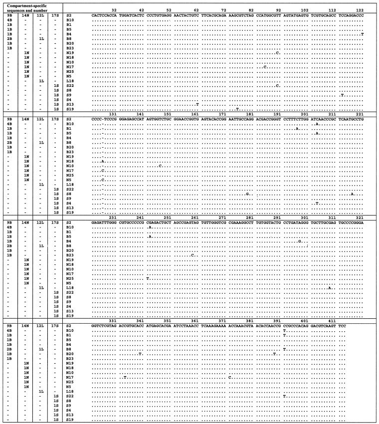
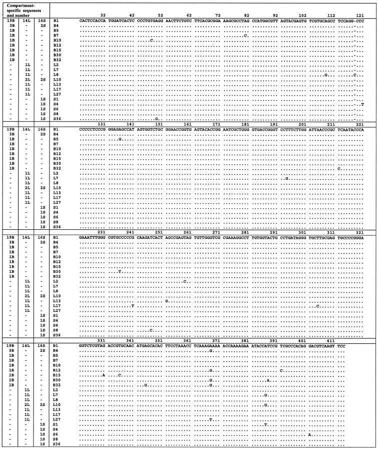
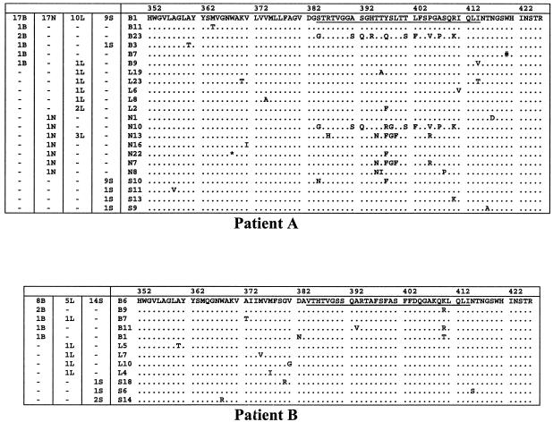
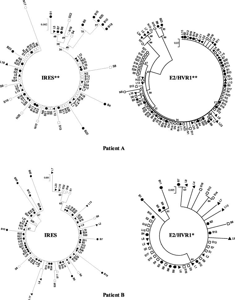
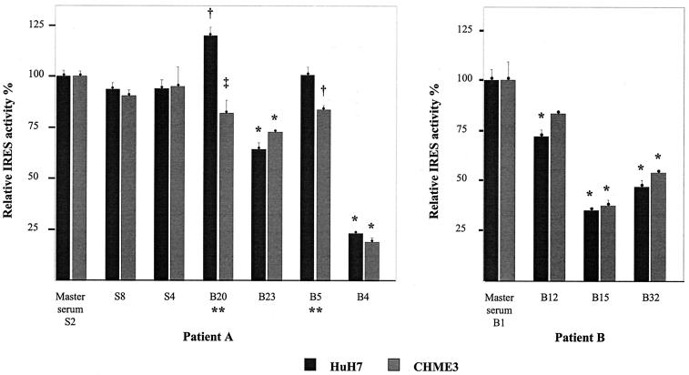
Reports of cerebral dysfunction in chronic hepatitis C virus (HCV) infection have led to the suggestion that HCV may infect the central nervous system (CNS). We used reverse transcription-PCR, cloning, and sequencing to define quasispecies for the HCV internal ribosomal entry site (IRES) and hypervariable region 1 (HVR1) in autopsy-derived brain, liver, lymph node, and serum samples. There was evidence of tissue compartmentalization of sequences in the brain in two patients, with between 24 and 55% of brain-derived IRES sequences absent from the serum, and significant phylogenetic and phenetic clustering of the brain and lymph node HVR1 sequences. The IRES initiates cap-independent translation of the viral polyprotein. Two unique brain-derived IRES mutations (C(204)-->A and G(243)-->A), which have previously been associated with lymphoid replication and altered translational efficiency in cell culture, were found in one patient. We used a dicistronic reporter vector to test whether brain-derived variants showed altered IRES-mediated translational efficiency, which might favor CNS infection. The translational efficiencies of the brain-derived IRES sequences were generally reduced compared to those of the master serum and liver sequences in rabbit reticulocyte cell lysates and two human cell lines, HuH7 (liver) and CHME3 (microglial). The C(204)-->A and G(243)-->A mutations showed preserved translational efficiency in HuH7 cells but reduced efficiency in CHME3 cells. Our data provide evidence that the CNS is a site of HCV replication, consistent with the recent demonstration of negative-strand HCV RNA in brain, and suggest that IRES polymorphisms may be important as a viral strategy of reduced translation to favor latency in the CNS.


Figures

FIG. 1.
FIG. 1.
Nucleotide alignment of IRES sequences amplified from brain, liver, serum, and lymph node for patient A. Sequences are compared to the master sequence, which was the same in each compartment. The numbers of sequences in each compartment are given in the four columns on the left. The identifier for each sequence is given in the fifth column. Nucleotide numbering as described by Choo et al. (7). B, brain; N, lymph node; L, liver; S, serum.
FIG. 2.
FIG. 2.
Nucleotide alignment of IRES sequences amplified from brain, liver, and serum in patient B. Sequences are compared to the master sequence, which was the same in each compartment. The numbers of sequences in each compartment are given in the three columns on the left. The identifier for each sequence is given in the fourth column. B, brain; N; L, liver; S, serum.
FIG. 3.
FIG. 3.
E2/HVR1 peptide alignments from serum and tissue compartments in patients A and B. These sequences are compared to the master sequence, which was the same in each compartment. The numbers of sequences in each compartment are given in the columns on the left. The identifier for each sequence is given before the sequence. B, brain; N, lymph node; L, liver; S, serum; *, stop codon; #, insertion of C at nucleotide position 1600. HVR1, comprising amino acids 384 to 414 is underlined in the master sequence.
FIG.4.
FIG.4.
Phylogenetic tree analyses of IRES and E2/HVR1 sequences from patients A and B. The trees representing the IRES nucleotide sequences were constructed by using the neighbor-joining algorithm and the Kimura two-parameter method in MEGA version 2.1. Trees representing the E2/HVR1 amino acid sequences were constructed by using the neighbor-joining algorithm and p-distances in MEGA version 2.1. Bootstrap values were derived from 500 resamplings of the original data. There was evidence of phenetic compartmentalization of the extrahepatic sequences where indicated (**, P < 0.001; *, P < 0.01 [Mantel test]). B, brain (•); N, lymph node (○); L, liver (▴); S, serum (□).
FIG. 5.
FIG. 5.
Translation efficiencies of HCV IRES variants in rabbit reticulocyte cell lysates from patients A and B. Translation efficiencies are shown relative to the master serum variant. The results shown represent the means obtained from at least three experiments for each IRES ± standard errors. *, P < 0.001.
FIG. 6.
FIG. 6.
Translation efficiencies of HCV IRES variants from patients A and B in HuH7 and CHME3 cell lines. Translation efficiencies are shown relative to the master serum variant. The results shown represent the means obtained from at least three experiments for each IRES ± standard errors. Differences were tested with analysis of variance and a Bonferroni correction. The following P values refer to comparisons with the master serum sequence: *, P < 0.001; †, P < 0.025; ‡, P = 0.05. There were statistical differences in translation efficiency in HuH7 and CHME3 cells for IRES variants B20 and B5 in patient A (**, P = 0.005).
FIG. 7.
FIG. 7.
Sequence and secondary structure of HCV IRES RNA (nucleotides 1 to 383, genotype 1b), showing the positions of mutations detected in patient A. Domains are numbered as described by Brown et al. (5). Sequence identifiers are shown in parentheses. Figure is modified from Honda et al. (18).

References

    1. Afonso, A. M., J. Jiang, F. Penin, C. Tareau, D. Samuel, M. A. Petit, H. Bismuth, E. Dussaix, and C. Feray. 1999. Nonrandom distribution of hepatitis C virus quasispecies in plasma and peripheral blood mononuclear cell subsets. J. Virol. 73:9213-9221. - PMC - PubMed
    1. Alter, M. J., H. S. Margolis, K. Krawczynski, F. N. Judson, A. Mares, W. J. Alexander, P. Y. Hu, J. K. Miller, M. A. Gerber, R. E. Sampliner, et al. 1992. The natural history of community-acquired hepatitis C in the United States. N. Engl. J. Med. 327:1899-1905. - PubMed
    1. Bouffard, P., P. H. Hayashi, R. Acevedo, N. Levy, and J. B. Zeldis. 1992. Hepatitis C virus is detected in a monocyte/macrophage subpopulation of peripheral blood mononuclear cells of infected patients. J. Infect. Dis. 166:1276-1280. - PubMed
    1. Bronowicki, J. P., M. A. Loriot, V. Thiers, Y. Grignon, A. L. Zignego, and C. Brechot. 1998. Hepatitis C virus persistence in human hematopoietic cells injected into SCID mice. Hepatology 28:211-218. - PubMed
    1. Brown, E. A., H. Zhang, L. H. Ping, and S. M. Lemon. 1992. Secondary structure of the 5′ nontranslated regions of hepatitis C virus and pestivirus genomic RNAs. Nucleic Acids Res. 20:5041-5045. - PMC - PubMed

Publication types

LinkOut - more resources