. 2004 May;78(10):5233-43.
doi: 10.1128/jvi.78.10.5233-5243.2004.

Analysis of host range phenotypes of primate hepadnaviruses by in vitro infections of hepatitis D virus pseudotypes

Affiliations

Analysis of host range phenotypes of primate hepadnaviruses by in vitro infections of hepatitis D virus pseudotypes

Azeneth Barrera et al. J Virol. 2004 May.

Abstract

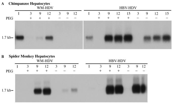
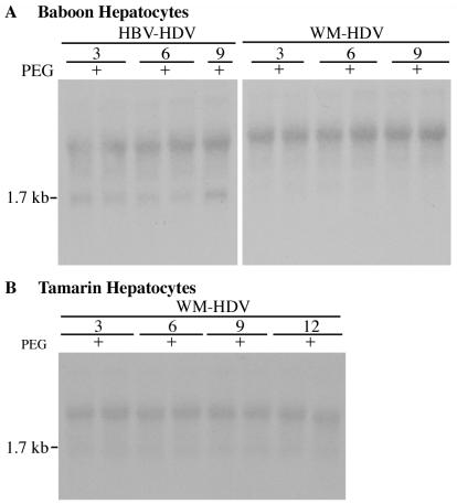
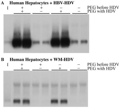
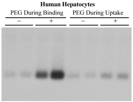
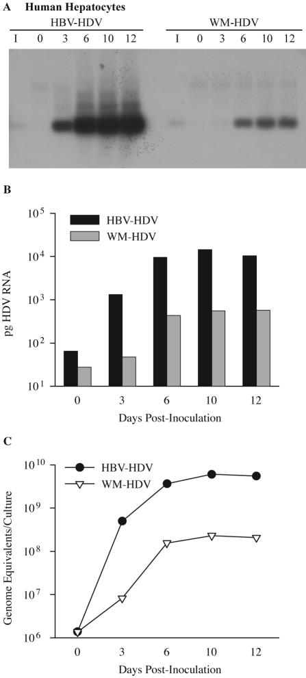
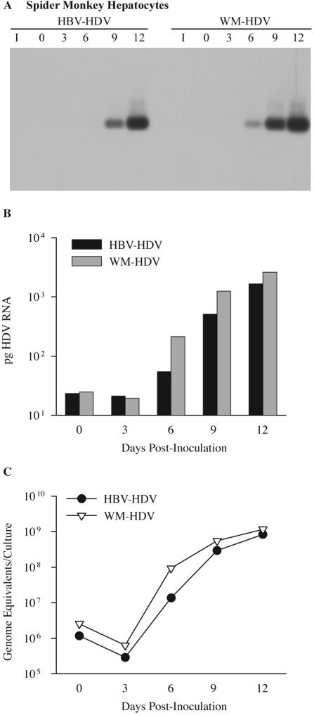
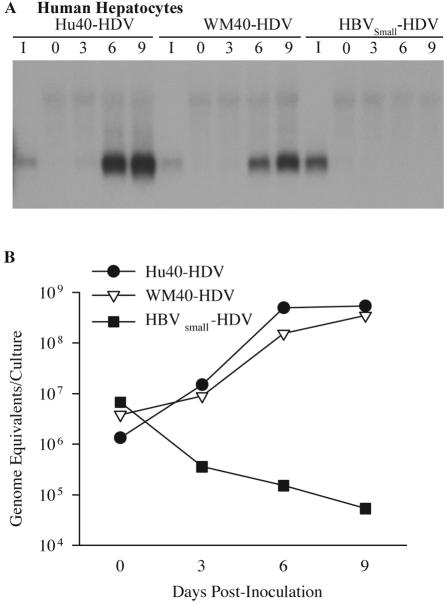
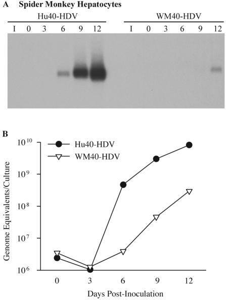
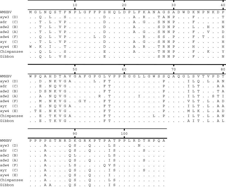
Hepatitis B virus (HBV) and woolly monkey hepatitis B virus (WMHBV) have natural host ranges that are limited to closely related species. The barrier for infection of primates seems to be at the adsorption and/or entry steps of the viral replication cycle, since a human hepatoma cell line is permissive for HBV and WMHBV replication following transfection of cloned DNA. We hypothesized that the HBV and WMHBV envelope proteins contain the principal viral determinants of host range. As previously shown by using the hepatitis D virus (HDV) system, recombinant HBV-HDV particles were infectious in chimpanzee as well as human hepatocytes. We extended the HDV system to include HDV particles pseudotyped with the WMHBV envelope. In agreement with the natural host ranges of HBV and WMHBV, in vitro infections demonstrated that HBV-HDV and WM-HDV particles preferentially infected human and spider monkey cells, respectively. Previous studies have implicated the pre-S1 region of the large (L) envelope protein in receptor binding and host range; therefore, recombinant HDV particles were pseudotyped with the hepadnaviral envelopes containing chimeric L proteins with the first 40 amino acids from the pre-S1 domain exchanged between HBV and WMHBV. Surprisingly, addition of the human amino terminus to the WMHBV L protein increased infectivity on spider monkey hepatocytes but did not increase infectivity for human hepatocytes. Based upon these data, we discuss the possibility that the L protein may be comprised of two domains that affect infectivity and that sequences downstream of residue 40 may influence host range and receptor binding or entry.


Figures

FIG. 1.
FIG. 1.
WM-HDV and HBV-HDV infection of primary hepatocytes in the presence or absence of PEG. Chimpanzee (A) and spider monkey (B) primary hepatocytes were inoculated with WM-HDV and HBV-HDV particles in the presence or absence of 5% PEG 8000. Cultures were harvested on days 3, 9, and 12 postinoculation (day 15 included for cells infected with HBV-HDV in panel A), and 5 μg of total cell RNA (approximately 15% of RNA from a 35-mm dish) was analyzed by Northern blot hybridization using a Riboprobe for HDV genomic RNA. RNA extracted from the equivalent of 5% of the inocula was analyzed under the same conditions (lane I). The size of HDV genomic RNA (in kilobases) is indicated.
FIG. 2.
FIG. 2.
Lack of infection of baboon and tamarin hepatocytes with HBV-HDV and WM-HDV. Cultures of baboon (A) and tamarin (B) hepatocytes were inoculated in duplicate with HBV-HDV (A) and WM-HDV (A and B) in the presence of 5% PEG. Cultures were harvested on days 3, 6, and 9 postinoculation, and total cellular RNA (5 μg) was analyzed by Northern blot hybridization using a Riboprobe for HDV genomic RNA. RNA extracted from 10% of the inocula was analyzed under the same conditions (lane I). The size of HDV genomic RNA (in kilobases) is indicated.
FIG. 3.
FIG. 3.
Requirement for simultaneous exposure of hepatocytes to PEG and virus for enhanced infection. Human hepatocytes were infected in duplicate with HBV-HDV (A) and WM-HDV (B) with or without prior exposure to PEG and with or without exposure to PEG during virus incubation. Preexposure to PEG (PEG before HDV, +/−) consisted of incubation in the presence of 5% PEG for 6 h at 37°C and then the removal of PEG by washing. Exposure to PEG plus virus (PEG with HDV, +/−) followed the standard protocol for exposure to virus in the presence or absence of 5% PEG. Cultures were harvested on day 9 postinoculation, and total cellular RNA (5 μg) was analyzed by Northern blot hybridization using a Riboprobe for HDV genomic RNA. RNA extracted from 10% of the inocula was analyzed under the same conditions (lane I).
FIG. 4.
FIG. 4.
PEG enhances virus binding but not postbinding events. PEG During Binding: HBV-HDV particles were allowed to adsorb onto human hepatocytes for 2 h at 4°C in the presence (+) or absence (−) of 5% PEG; the cultures were then washed and incubated overnight at 37°C in the absence of PEG. PEG During Uptake: virus was allowed to adsorb onto hepatocytes for 2 h at 4°C in the absence of PEG; the cultures were washed and then incubated overnight at 37°C in the presence (+) or absence (−) of PEG. Cultures were harvested on day 12 postinoculation, and total cellular RNA (5 μg) was analyzed by Northern blot hybridization using a Riboprobe for HDV genomic RNA. RNA extracted from 10% of the inocula was analyzed under the same conditions (lane I).
FIG. 5.
FIG. 5.
Infectivity of HDV pseudotypes of HBV and WMHBV in human hepatocytes. (A) Human hepatocytes were infected with HBV-HDV or WM-HDV particles. Cultures were harvested on days 0, 3, 6, 10, and 12 postinoculation, and total cellular RNA (5 μg) was analyzed by Northern blot hybridization using a Riboprobe for HDV genomic RNA. RNA extracted from 10% of the inocula was analyzed under the same conditions (lane I). (B) Autoradiographs from panel A were scanned with a phosphorimager. The amount of HDV RNA is expressed in picograms per culture and was derived by linear regression analysis of HDV RNA standards loaded on the same gel. (C) Levels of HDV RNA from the same cultures analyzed in panels A and B were quantified by TaqMan RT-PCR and expressed as genomic equivalents per culture.
FIG. 6.
FIG. 6.
Infectivity of HDV pseudotypes of HBV and WMHBV in spider monkey hepatocytes. (A) Spider monkey hepatocytes were infected with HBV-HDV and WM-HDV particles. Cultures were harvested on days 0, 3, 6, 9, and 12 postinoculation, and total cellular RNA was analyzed as described in the legend for Fig. 5. (B) Autoradiographs from panel A were scanned with a phosphorimager. The amount of HDV RNA is expressed in picograms per culture and was derived by linear regression analysis of HDV RNA standards loaded on the same gel. (C) Levels of HDV RNA from the same cultures analyzed in panels A and B were quantified by TaqMan RT-PCR and expressed as genomic equivalents per culture.
FIG. 7.
FIG. 7.
Infectivity of HDV pseudotypes with chimeric HBV and WMHBV L proteins in human hepatocytes. (A) Human hepatocytes were infected with Hu40-HDV and WM40-HDV particles. Cultures were also inoculated with HDV pseudotypes containing only the HBV S protein (HBVSmall-HDV) in the presence of 5% PEG. Cultures were harvested on days 0, 3, 6, and 9 postinoculation, and total cellular RNA was analyzed as described in the legend for Fig. 5. (B) Levels of HDV RNA from the same cultures analyzed in panel A were quantified by TaqMan RT-PCR and expressed as genomic equivalents per culture.
FIG. 8.
FIG. 8.
Infectivity of HDV pseudotypes with chimeric HBV and WMHBV L proteins in spider monkey hepatocytes. (A) Spider monkey hepatocytes were infected with Hu40-HDV and WM40-HDV particles. Cultures were harvested on days 0, 3, 6, 9, and 12 postinoculation, and total cellular RNA was analyzed as described in the legend for Fig. 5. (B) Levels of HDV RNA from the same cultures analyzed in panel A were quantified by TaqMan RT-PCR and expressed as genomic equivalents per culture.
FIG. 9.
FIG. 9.
Alignment of HBV pre-S1 domains. The amino acid sequences of the pre-S1 domains for HBV genotypes A to F are displayed along with the sequences for WMHBV and HBV isolates from chimpanzees and gibbons. The GenBank numbers for the isolates are AY226578 (WMHBV), V01460 and J02203 (ayw3-D), L08805 (adr-C), M54923 (adw2-B), X02763 (adw2-A), X75663 (adw4-F), X04615 (ayr-C), X75664 (ayw4-E), D00220 (chimpanzee), and U46935 (gibbon).

References

    1. Barrera, A., and R. E. Lanford. 2004. Infection of primary chimpanzee hepatocytes with recombinant hepatitis D virus particles: a surrogate model for hepatitis B virus. Methods Mol. Med. 95:131-142. - PubMed
    1. Beames, B., and R. E. Lanford. 1993. Carboxy-terminal truncations of the HBV core protein affect capsid formation and size of the encapsidated HBV RNA. Virology 194:597-607. - PubMed
    1. Bonino, F., K. H. Heermann, M. Rizzetto, and W. H. Gerlich. 1986. Hepatitis delta virus: protein composition of delta antigen and its hepatitis B virus-derived envelope. J. Virol. 58:945-950. - PMC - PubMed
    1. Bonino, F., B. Hoyer, J. W.-K. Shih, M. Rizzetto, R. H. Purcell, and J. L. Gerin. 1984. Delta hepatitis agent: structural and antigenic properties of the delta-associated particle. Infect. Immun. 43:1000-1005. - PMC - PubMed
    1. Breiner, K. M., S. Urban, and H. Schaller. 1998. Carboxypeptidase D (gp180), a Golgi-resident protein, functions in the attachment and entry of avian hepatitis B viruses. J. Virol. 72:8098-8104. - PMC - PubMed

Publication types