. 2004 May;78(10):5258-69.
doi: 10.1128/jvi.78.10.5258-5269.2004.

Role of transmembrane domain and cytoplasmic tail amino acid sequences of influenza a virus neuraminidase in raft association and virus budding

Affiliations

Role of transmembrane domain and cytoplasmic tail amino acid sequences of influenza a virus neuraminidase in raft association and virus budding

Subrata Barman et al. J Virol. 2004 May.

Abstract

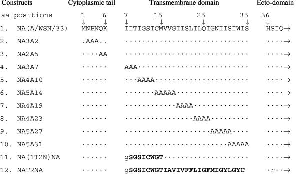
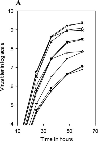
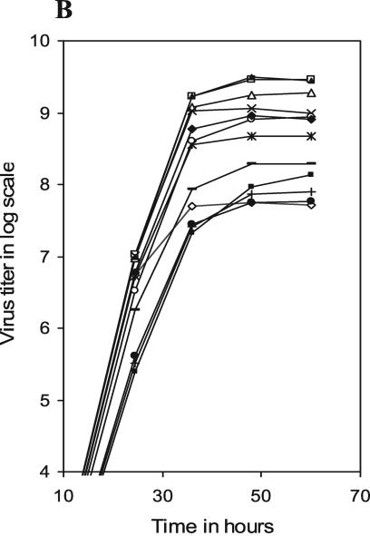
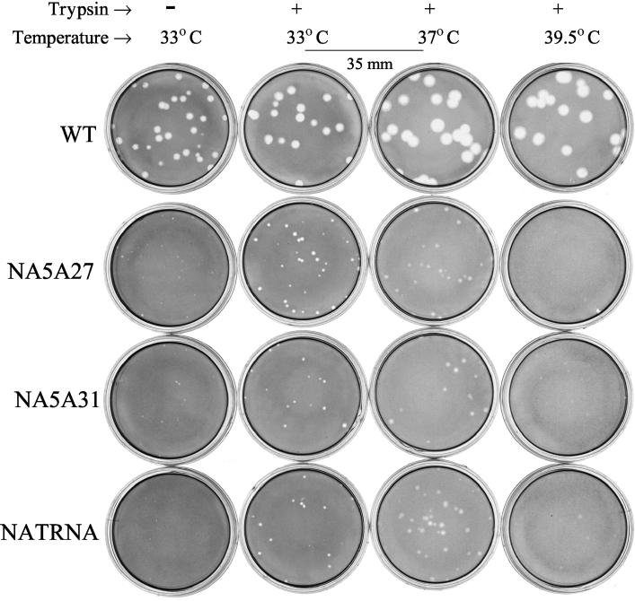
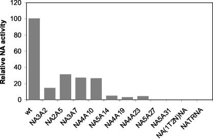
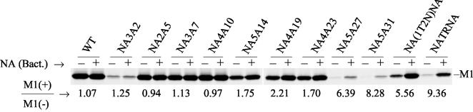
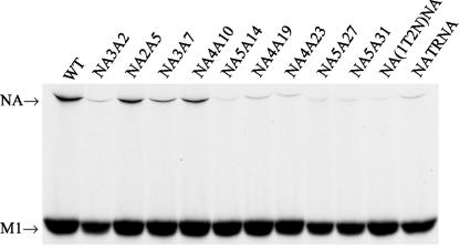
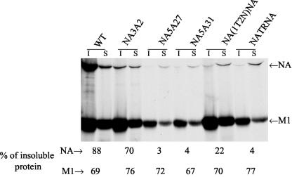
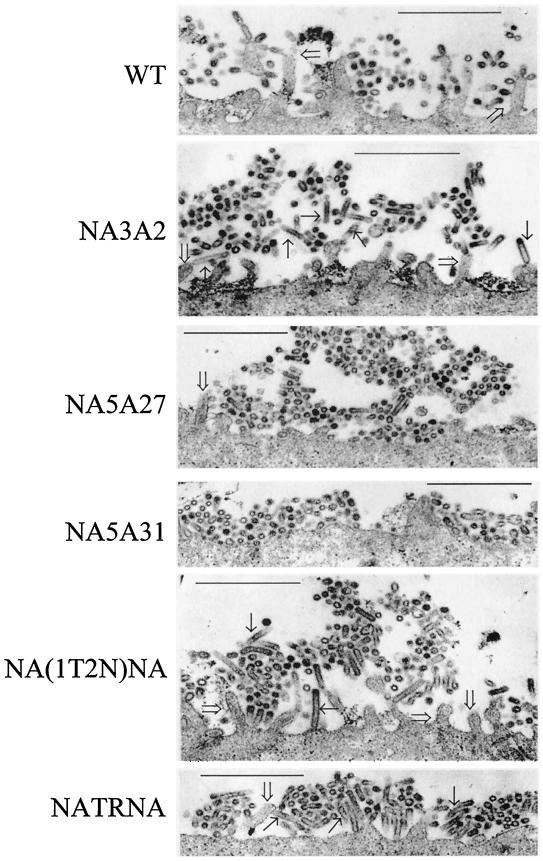
Influenza virus neuraminidase (NA), a type II transmembrane glycoprotein, possesses receptor-destroying activity and thereby facilitates virus release from the cell surface. Among the influenza A viruses, both the cytoplasmic tail (CT) and transmembrane domain (TMD) amino acid sequences of NA are highly conserved, yet their function(s) in virus biology remains unknown. To investigate the role of amino acid sequences of the CT and TMD on the virus life cycle, we systematically mutagenized the entire CT and TMD of NA by converting two to five contiguous amino acids to alanine. In addition, we also made two chimeric NA by replacing the CT proximal one-third amino acids of the NA TMD [NA(1T2N)NA] and the entire NA TMD (NATRNA) with that of human transferrin receptor (TR) (a type II transmembrane glycoprotein). We rescued transfectant mutant viruses by reverse genetics and examined their phenotypes. Our results show that all mutated and chimeric NAs could be rescued into transfectant viruses. Different mutants showed pleiotropic effects on virus growth and replication. Some mutants (NA2A5, NA3A7, and NA4A10) had little effect on virus growth while others (NA3A2, NA5A27, and NA5A31) produced about 50- to 100-fold-less infectious virus and still some others (NA5A14, NA4A19, and NA4A23) exhibited an intermediate phenotype. In general, mutations towards the ectodomain-proximal sequences of TMD progressively caused reduction in NA enzyme activity, affected lipid raft association, and attenuated virus growth. Electron microscopic analysis showed that these mutant viruses remained aggregated and bound to infected cell surfaces and could be released from the infected cells by bacterial NA treatment. Moreover, viruses containing mutations in the extreme N terminus of the CT (NA3A2) as well as chimeric NA containing the TMD replaced partially [NA(1T2N)NA] or fully (NATRNA) with TR TMD caused reduction in virus growth and exhibited the morphological phenotype of elongated particles. These results show that although the sequences of NA CT and TMD per se are not absolutely essential for the virus life cycle, specific amino acid sequences play a critical role in providing structural stability, enzyme activity, and lipid raft association of NA. In addition, aberrant morphogenesis including elongated particle formation of some mutant viruses indicates the involvement of NA in virus morphogenesis and budding.


Figures

FIG. 1.
FIG. 1.
Schematic presentation of NA mutants. Sets of 2 to 5 aa in the NA CT or TMD were replaced with alanines. Chimeric NA TMDs [NA(1T2N)NA and NATRNA] were constructed by swapping either the first 9 aa or the entire NA TMD with that of the TR TMD. ·, amino acids from NA sequences. Amino acids shown in boldface type are from TR sequences. Amino acids in lowercase type are due to created restriction enzyme sites. Note that the TR TMD contains 28 aa instead of the 29 aa of the NA TMD. Numbers above amino acid sequences represent amino acid positions (indicated by ↓) of the WT NA peptide.
FIG. 2.
FIG. 2.
Growth curves of different mutant viruses. MDCK cell monolayers were infected with viruses at an MOI of 0.001 and maintained at 33°C in VGM without (A) or with (B) 0.5 μg of TPCK-treated trypsin/ml. At different time points p.i., aliquots of culture supernatant were collected and titrated for infectious viruses by PFU assay. ▴, WT; ⋄, NA3A2; □, NA2A5; Δ, NA3A7; ×, NA4A10; Ж, NA5A14; ○, NA4A19; ♦, NA4A23; •, NA5A27; +, NA5A31; −, NA(1T2N)TR; ▪, NATRNA. Results represent averages of the results from four independent experiments.
FIG. 2.
FIG. 2.
Growth curves of different mutant viruses. MDCK cell monolayers were infected with viruses at an MOI of 0.001 and maintained at 33°C in VGM without (A) or with (B) 0.5 μg of TPCK-treated trypsin/ml. At different time points p.i., aliquots of culture supernatant were collected and titrated for infectious viruses by PFU assay. ▴, WT; ⋄, NA3A2; □, NA2A5; Δ, NA3A7; ×, NA4A10; Ж, NA5A14; ○, NA4A19; ♦, NA4A23; •, NA5A27; +, NA5A31; −, NA(1T2N)TR; ▪, NATRNA. Results represent averages of the results from four independent experiments.
FIG. 3.
FIG. 3.
Plaque morphologies of different mutant viruses. MDCK cell monolayers (35-mm-diameter dish) were infected with serially diluted viruses and maintained at different temperatures in agarose overlay medium with (+) or without (−) TPCK-treated trypsin (0.5 μg/ml). At 60 h p.i., the agarose overlay medium was removed, stained for 1 min with crystal violet, and washed with PBS.
FIG. 4.
FIG. 4.
Sialidase activity of mutant viruses. Sialidase activity was determined with the 2′-(4-methylumbelliferyl)-α-d-N-acetylneuraminic acid substrate as described in Materials and Methods. Specific activity was calculated by normalizing NA activity to the level of virus particles, as determined by immunoblotting viral proteins (separated by SDS-PAGE) with antibody against M1, followed by densitometric analysis. Relative NA activity, taking that of WT virus as 100%, was calculated from the results from three independent experiments, with an average variation of less than 10%.
FIG. 5.
FIG. 5.
Effect of bacterial NA on virus release. Virus-infected MDCK cells were metabolically labeled (at 5 h p.i.) with 300 μCi of 35S-protein labeling mix in 2:8 VGM for 9 h. Two hours before collection, virus-infected cells were treated (+) or mock treated (−) with bacterial NA (10 mU/ml). At 14 h p.i., released virus particles in the supernatant were collected and clarified by low-speed centrifugation and virions were purified and pelleted by ultracentrifugation through a 25% sucrose cushion. Pellets were dissolved in TNE buffer. Viral proteins were analyzed directly by SDS-PAGE, autoradiographed, and quantified. The increase in virus release after NA treatment was determined by using the M1 band. The average value of M1(+)/M1(−) was calculated from the results from three to five independent experiments, with a variation of less than 10%.
FIG. 6.
FIG. 6.
NA protein incorporation in virions. Virus-infected MDCK cells were labeled at 4 h p.i. for 12 h with 200 μCi of 35S-protein labeling mix in 2:8 VGM. At 16 h p.i., medium was harvested and clarified by low-speed centrifugation and virions were pelleted by ultracentrifugation through a 25% sucrose cushion. Labeled virion lysates were immunoprecipitated with the respective antibodies; proteins were eluted, treated with PNGase F for 3 h, analyzed by SDS-PAGE, and autoradiographed.
FIG. 7.
FIG. 7.
Triton X-100 insolubility of cell surface proteins. At 4 h p.i., influenza virus-infected (2.5 h p.i. for VSV-infected cells) MDCK cells were metabolically labeled for 60 min with 300 μCi of 35S-protein labeling mix/ml and chased for 90 min. Cell surface proteins were biotinylated and extracted with 1% Triton X-100 on ice for 10 min. Triton X-100-soluble (S) and -insoluble (I) fractions of surface biotinylated proteins were immunoprecipitated with specific antibodies for NA, HA, or VSV G proteins, analyzed by SDS-PAGE, autoradiographed, and quantified. The percentage of Triton X-100-insoluble protein was calculated from the results from three to five independent experiments, with average variation of less than 10%. *, the Triton X-100 insolubility of HA was calculated with both HA0 and HA1 bands.
FIG. 8.
FIG. 8.
Triton X-100 insolubility of different proteins in virions. 35S-labeled virions were prepared as described in the legend to Fig. 6 and treated with 0.1% Triton X-100 on ice for 10 min, soluble (S) and insoluble (I) fractions were immunoprecipitated with the respective antibodies, and proteins were eluted, treated with PNGase F for 3 h, analyzed by SDS-PAGE, autoradiographed, and quantified. The percentage of Triton X-100-insoluble protein was calculated from the results from three to five independent experiments, with an average variation of less than 10%.
FIG. 9.
FIG. 9.
Budding of viruses by thin-section electron microscopy. MDCK cells grown on polycarbonate filters were infected with different viruses at an MOI of 3.0. At 12 h p.i., infected cell monolayers were cross-linked in 2% glutaraldehyde and postfixed with 1% OsO4. Filters were embedded in Epon, and 60-nm-thick sections were stained with uranyl acetate and lead citrate and examined with a transmission electron microscope. Bar, 1 μm. →, elongated virus particles; ⇒, villi.

References

    1. Air, G. M., and W. G. Laver. 1989. The neuraminidase of influenza virus. Proteins 6:341-356. - PubMed
    1. Ali, A., R. T. Avalos, E. Ponimaskin, and D. P. Nayak. 2000. Influenza virus assembly: effect of influenza virus glycoproteins on the membrane association of M1 protein. J. Virol. 74:8709-8719. - PMC - PubMed
    1. Barman, S., L. Adhikary, Y. Kawaoka, and D. P. Nayak. 2003. Influenza A virus hemagglutinin containing basolateral localization signal does not alter the apical budding of a recombinant influenza A virus in polarized MDCK cells. Virology 305:138-152. - PubMed
    1. Barman, S., A. Ali, E. K.-W. Hui, L. Adhikary, and D. P. Nayak. 2001. Transport of viral proteins to the apical membranes and interaction of matrix protein with glycoproteins in the assembly of influenza viruses. Virus Res. 77:61-69. - PubMed
    1. Barman, S., and D. P. Nayak. 2000. Analysis of the transmembrane domain of influenza virus neuraminidase, a type II transmembrane glycoprotein, for apical sorting and raft association. J. Virol. 74:6538-6545. - PMC - PubMed

Publication types

LinkOut - more resources