

Review
doi: 10.1379/1466-1268(2003)008<0309:mfrohf>2.0.co;2.
Mechanisms for regulation of Hsp70 function by Hsp40
Affiliations
- PMID: 15115283
- PMCID: PMC514902
- DOI: 10.1379/1466-1268(2003)008<0309:mfrohf>2.0.co;2
Item in Clipboard
Review
Mechanisms for regulation of Hsp70 function by Hsp40
Cell Stress Chaperones.
2003 Winter.
Abstract
The Hsp70 family members play an essential role in cellular protein metabolism by acting as polypeptide-binding and release factors that interact with nonnative regions of proteins at different stages of their life cycles. Hsp40 cochaperone proteins regulate complex formation between Hsp70 and client proteins. Herein, literature is reviewed that describes the mechanisms by which Hsp40 proteins interact with Hsp70 to specify its cellular functions.
Figures
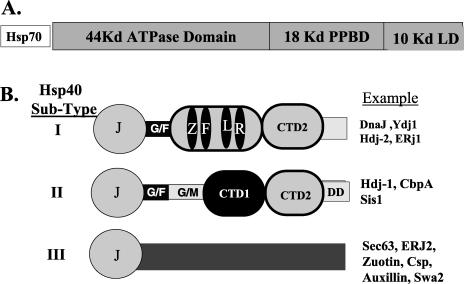
Schematic diagrams of Hsp70 and members of the Hsp40 family. (A) The organization of Hsp70 into different subdomains. The 44-kDa adenosine triphosphatase (ATPase) domain represents a 44-kDa amino terminal fragment of Hsp70 that contains the ATP-binding site and retains ATPase activity. The 18-kDa polypeptide-binding domain (PPBD) represents an internal fragment of Hsp70 that functions as the PPD. The 10-kDa lid domain (LD) is a C-terminal fragment that is proposed to function as an LD that covers the PPBD and serves as a site for the binding of cochaperones. (B) Domain structure of different Hsp40 subtypes. J, J-domain; G/F, glycine-and phenylalanine-rich region; ZFLR, zinc finger–like region; G/M, glycine/methioine-rich region; CTDI, carboxyl-terminal domain I; CTDII, is carboxyl terminal domain II; DD, the dimerization domain. The examples represent Hsp40 proteins from E coli, S cerevisiae, and H sapiens
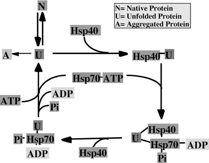
A proposed model for Hsp40-dependent polypeptide binding and release by Hsp70. Hsp40 proteins form complexes with unfolded or nonnative proteins to prevent their aggregation. Hsp40 then delivers the unfolded protein to Hsp70. Stable Hsp70-protein complexes are then formed by a mechanism that involves Hsp40 J-domain dependent conversion of Hsp70-adenosine triphosphate (ATP) to Hsp70-adenosine diphosphate. Hsp70-protein complexes dissociate upon regeneration of Hsp70-ATP. Upon release from Hsp70, an unfolded polypeptide can fold, aggregate, or be rebound by Hsp40 and Hsp70

Structures of the Hsp40 J-domain and the Hsp70 adenosine triphosphatase (ATPase) domain. (A) Ribbon diagram of the nuclear magnetic resonance solution structure of residues 2–77 of E coli DnaJ. HPD denotes the position of the conserved HPD motif that is found in the J-domain of all Hsp40s. D in the HPD motif corresponds to D35 in DnaJ that was mutated in experiments that identified the J-domain–binding site in DnaK (see text for details). Gly78 denotes the end of the J-domain and the beginning of the glycine- and phenylalanine-rich region. (B) Ribbon diagram of the X-ray crystal structure of the 382-residue amino-terminal fragment of Hsp70 that retains ATPase activity. Glu175 is the conserved residue in Hsp70 that is represented in E coli DnaK (Hsp70) by Glu171. This residue is denoted because it was demonstrated to function in interdomain communication between the ATPase domain and the polypeptide-binding domain of Hsp70 (see text). Glu175 is also located near R167, which is found in an acidic cleft where J-domain binding to Hsp70 is proposed to occur (see text). Gly382 denotes the terminus of ATPase domain. β-Strands are shown in gold, and α-helices are in pink. The J-domain and Hsp70 ATPase fragment structures were from PDB files 1BQZ and 1HJO, respectively, and the images shown were generated with Rasmol
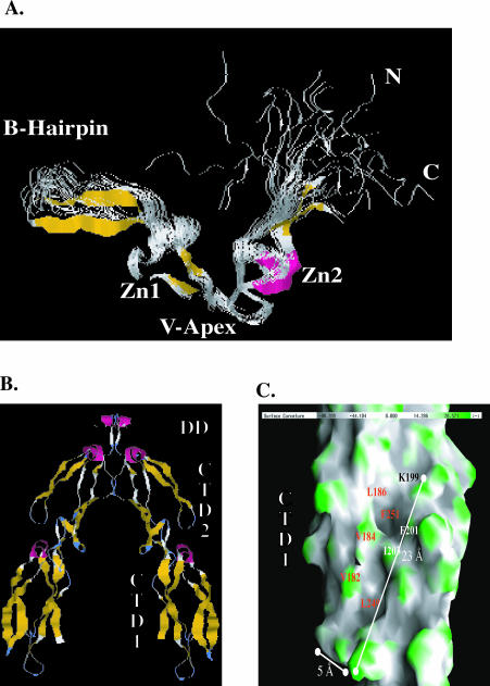
Structure of regions in type I and type II Hsp40s that are involved in chaperone function. (A) The nuclear magnetic resonance solution structure of the zinc finger–like region (ZFLR) from E coli DnaJ. A ribbon diagram depicting the nuclear magnetic resonance solution structure of a protein fragment that corresponds to Gly131 to Ser209 of E coli DnaJ. This image was rendered from PDB file 1EXK. β-Strands are shown in gold, and α-helices are in pink. Zn1 and Zn2 denote the 2 regions in the ZFLR where zinc is bound. N and C denote the position of resides 8 and 79 of DnaJ 131–209. This diagram represents 20 structures. (B) Ribbon diagram of the Sis1 (171–352) dimer. Sis1 (171–352) is truncated at the end of the glycine- and methionine-rich region (G/M) region, and this model shows residues 180–352. A and B represent the monomers that form the Sis1 (171–352) dimer, which has a 2-fold axis. Subdomains present in the Sis1 (171–352) monomer are labeled. Carboxyl-terminal domain I (CTDI) corresponds to residues 180–254. CTDII corresponds to residues 255–341. The Sis1 dimerization domain (DD) lies between residues 341 and 352. β-Strands are shown in gold, and α-helices are in pink. (C) An enlarged view of the surface of CTDI as depicted in monomer B of (B). The surface shown depicts contours on CTDI with concave areas in gray and convex areas in green. A surface hydrophobic groove that contains 2 shallow depressions is visible. Solvent-exposed residues that line the depression are denoted. F251 and L249 form the base of the individual depressions. Panels in (B) and (C) were rendered from PDB file 1C3G with Rasmol and GRASP software packages, respectively
References
-
- Banecki B, Liberek K, Wall D, Wawrzynow A, Georgopoulos C, Bertoli E, Tanfani F, Zylicz M. Structure-function analysis of the zinc finger region of the DnaJ molecular chaperone. J Biol Chem. 1996;271:14840–14848. - PubMed
-
- Bischofberger P, Han W, Feifel B, Schonfeld HJ, Christen P. d-Peptides as inhibitors of the DnaK/DnaJ/GrpE chaperone system. J Biol Chem. 2003;278:19044–19047. - PubMed
-
- Bukau B, Horwich AL. The Hsp70 and Hsp60 chaperone machines. Cell. 1998;92:351–366. - PubMed
Publication types
MeSH terms
Substances
Grants and funding
LinkOut - more resources
Full Text Sources
Other Literature Sources