. 2004 Sep 1;382(Pt 2):725-31.
doi: 10.1042/BJ20040413.

Deletion of Ser-171 causes inactivation, proteasome-mediated degradation and complete deficiency of human transaldolase

Affiliations

Deletion of Ser-171 causes inactivation, proteasome-mediated degradation and complete deficiency of human transaldolase

Craig E Grossman et al. Biochem J. .

Abstract

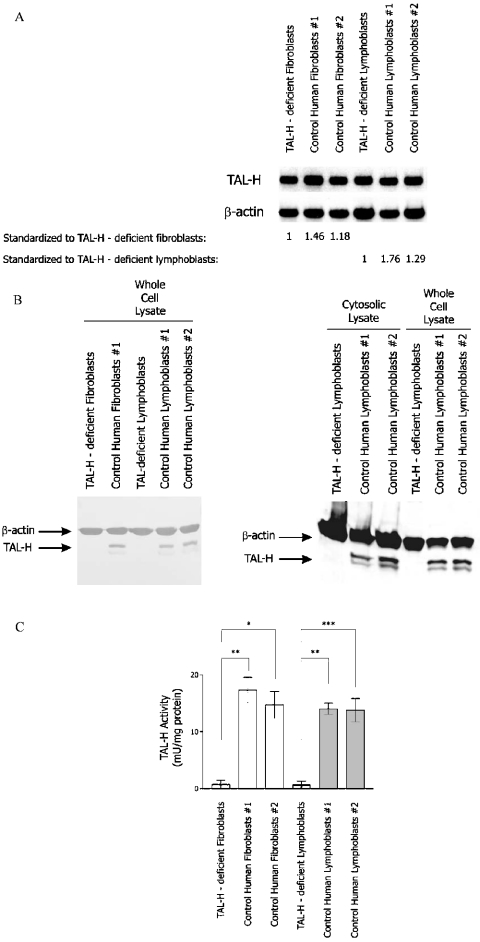
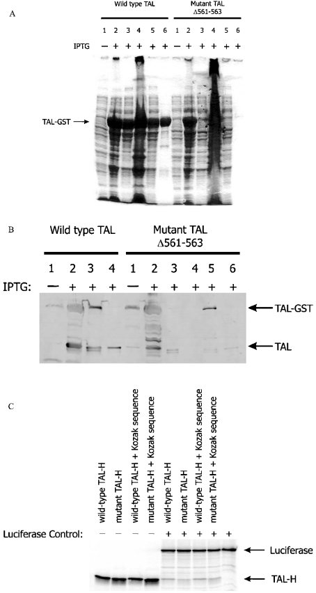
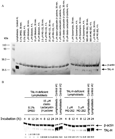
Homozygous deletion of three nucleotides coding for Ser-171 (S171) of TAL-H (human transaldolase) has been identified in a female patient with liver cirrhosis. Accumulation of sedoheptulose 7-phosphate raised the possibility of TAL (transaldolase) deficiency in this patient. In the present study, we show that the mutant TAL-H gene was effectively transcribed into mRNA, whereas no expression of the TALDeltaS171 protein or enzyme activity was detected in TALDeltaS171 fibroblasts or lymphoblasts. Unlike wild-type TAL-H-GST fusion protein (where GST stands for glutathione S-transferase), TALDeltaS171-GST was solubilized only in the presence of detergents, suggesting that deletion of Ser-171 caused conformational changes. Recombinant TALDeltaS171 had no enzymic activity. TALDeltaS171 was effectively translated in vitro using rabbit reticulocyte lysates, indicating that the absence of TAL-H protein in TALDeltaS171 fibroblasts and lymphoblasts may be attributed primarily to rapid degradation. Treatment with cell-permeable proteasome inhibitors led to the accumulation of TALDeltaS171 in whole cell lysates and cytosolic extracts of patient lymphoblasts, suggesting that deletion of Ser-171 led to rapid degradation by the proteasome. Although the TALDeltaS171 protein became readily detectable in proteasome inhibitor-treated cells, it displayed no appreciable enzymic activity. The results suggest that deletion of Ser-171 leads to inactivation and proteasome-mediated degradation of TAL-H. Since TAL-H is a regulator of apoptosis signal processing, complete deficiency of TAL-H may be relevant for the pathogenesis of liver cirrhosis.


Figures

Figure 1
Figure 1. Measurement of TAL expression by Northern- and Western-blot analyses and enzyme activity assays
(A) Northern-blot analysis of steady-state TAL mRNA levels in TALΔS171 and control fibroblasts and lymphoblasts. Total RNA (10 μg) from TALΔS171 (TAL-H-deficient) and control fibroblasts and lymphoblasts were separated on 1% agarose gel, transferred on to a nylon membrane and hybridized to TAL cDNA probe 4/1 [9] or human β-actin probe [10], used as an internal control. The relative abundance of TAL transcripts in control cells with respect to TALΔS171 fibroblasts and lymphoblasts set at 1.0 is shown under columns 2 and 3. (B) Western-blot analysis of TAL expression in whole cell extracts (2×10⁵ cells/lane) and cytosolic protein lysates (20 μg/lane) of TALΔS171 and control lymphoblasts. TAL was detected with antibody 170. Actin levels were monitored as loading control. (C) Measurement of TAL enzyme activity in TALΔS171 and control fibroblasts and lymphoblasts. Results are expressed as the means±S.E.M. for five or more independent experiments. *P<0.05, **P<0.01 and ***P<0.001.
Figure 2
Figure 2. Production of mutant TAL by prokaryotic expression vectors and in vitro translation
(A) Prokaryotic expressions of wild-type and mutant TALΔ561–563 cDNAs. The TAL-H protein was expressed as a fusion protein with GST encoded by pGEX-2T plasmid vector [11]. Maximal expression of the recombinant fusion protein was obtained after stimulation with 1 mM IPTG for 2 h. TAL-H–GST fusion protein was affinity-purified through binding of GST to glutathione-coated agarose beads. Protein lysates were analysed on 12% SDS/polyacrylamide gel. Lane 1, whole cell lysates from unstimulated cells; lane 2, whole cell lysates from IPTG-stimulated cells; lane 3, supernatant of IPTG-stimulated cells disrupted by freezing and thawing performed three times; lane 4, pellet of IPTG-stimulated cells disrupted by freezing and thawing performed three times; lane 5, supernatant of IPTG-stimulated cells incubated with GSH-coated agarose beads; lane 6, GSH-coated agarose beads pelleted after exposure to IPTG-stimulated cell supernatant. (B) Western-blot detection of solubilized, affinity-purified and thrombin-cleaved recombinant TAL–GST fusion proteins. IPTG-stimulated cells were disrupted in the absence or presence of 1.5% N-laurylsarcosine, 2% Tween 20 and 4% Triton X-100, and supernatants were incubated with GSH-coated agarose beads. Subsequently, beads were washed six times in 1 ml of PBS, digested overnight with thrombin in 500 μl of PBS and tested for the presence of TAL by Western blotting using antibody 170. Lane 1, whole cell lysate from unstimulated cells; lane 2, whole cell lysate from IPTG-stimulated cells; lane 3, GSH-coated agarose beads exposed to the supernatant of IPTG-stimulated cells disrupted by freezing and thawing performed three times; lane 4, supernatant of thrombin-treated agarose beads previously exposed to the supernatant of IPTG-stimulated cells disrupted by freezing and thawing three times; 5, GSH-coated agarose beads exposed to the supernatant of IPTG-stimulated cells disrupted by freezing and thawing three times in the presence of 1.5% N-laurylsarcosine, 2% Tween 20 and 4% Triton X-100; lane 6, supernatant of thrombin-treated agarose beads previously exposed to the supernatant of IPTG-stimulated cells disrupted by freezing and thawing three times in the presence of 1.5% N-laurylsarcosine, 2% Tween 20 and 4% Triton X-100. (C) In vitro translation of wild-type and mutant TALΔ561–563 RNA by rabbit reticulocyte lysates. For generating the [35S]methionine-labelled product, 1 μg of pCMVTNT-based wild-type or mutant TAL-H cDNA was mixed with nuclease-free rabbit reticulocyte lysate, T7 RNA polymerase, amino acids minus methionine and 20 μCi of L-[35S]methionine and incubated at 30 °C for 90 min. As indicated, firefly luciferase-encoding control plasmid driven by the T7 RNA polymerase promoter was added to the reaction mixture.
Figure 3
Figure 3. Conformational changes elicited by the deletion of Ser-171 of TAL
(A) Overlay of three-dimensional models of TAL-H and TALΔS171 reveals remarkably similar global structures. (B) Phe-171 of TALΔS171 is positioned, by 3.66 Å, closer to the centre of the α/β-barrel when compared with Phe-172 of TAL-H.
Figure 4
Figure 4. Effects of cell-permeable proteasome inhibitors on TAL protein levels in TALΔS171 lymphoblasts
(A) Proteasome inhibitors gliotoxin, MG-132, clasto-lactacystin β-lactone, epoxomycin and MG-262 were dissolved in DMSO and added to 106 cells. When used as a solvent for proteasome inhibitors, the final concentration of DMSO was less than 0.01%. After incubation for 30 min or 4 h, protein lysates from whole cell extracts were analysed by Western blotting. TAL was detected with antibody 170 and human β-actin was monitored with the monoclonal antibody 1501R. (B) Effects of the proteasome inhibitors clasto-lactacystin β-lactone and MG-262 on accumulation of TAL-H in TALΔS171 lymphoblasts. Using automated densitometry, TAL-H expression levels of proteasome-treated TALΔS171 lymphoblasts were normalized to internal actin levels and are compared with cells exposed to 0.1% DMSO alone for the same length of time.

References

    1. Mayes P. A. The pentose phosphate pathway and other pathways of hexose metabolism. In: Murray R. K., Granner D. K., Mayes P. A., Rodwell V. W., editors. Harper's Biochemistry. Norwalk, CT: Appleton and Lange; 1993. pp. 201–211.
    1. Cooper R. A., Bunn H. F. Hemolytic anemias. In: Wilson J. D., Braunwald E., Isselbacher K. J., Petersdorf R. G., Martin J. B., Fauci A. S., Root R. K., editors. Harrison's Principles of Internal Medicine. New York: McGraw-Hill; 1991. pp. 1531–1543.
    1. Banki K., Hutter E., Colombo E., Gonchoroff N. J., Perl A. Glutathione levels and sensitivity to apoptosis are regulated by changes in transaldolase expression. J. Biol. Chem. 1996;271:32994–33001. - PubMed
    1. Banki K., Hutter E., Gonchoroff N. J., Perl A. Molecular ordering in HIV-induced apoptosis: oxidative stress, activation of caspases, and cell survival are regulated by transaldolase. J. Biol. Chem. 1998;273:11944–11953. - PubMed
    1. Banki K., Hutter E., Gonchoroff N., Perl A. Elevation of mitochondrial transmembrane potential and reactive oxygen intermediate levels are early events and occur independently from activation of caspases in Fas signaling. J. Immunol. 1999;162:1466–1479. - PMC - PubMed

Publication types

MeSH terms