. 2004 Apr 28:2:20.
doi: 10.1186/1477-7827-2-20.

Origin of germ cells and formation of new primary follicles in adult human ovaries

Affiliations

Origin of germ cells and formation of new primary follicles in adult human ovaries

Antonin Bukovsky et al. Reprod Biol Endocrinol. .

Abstract

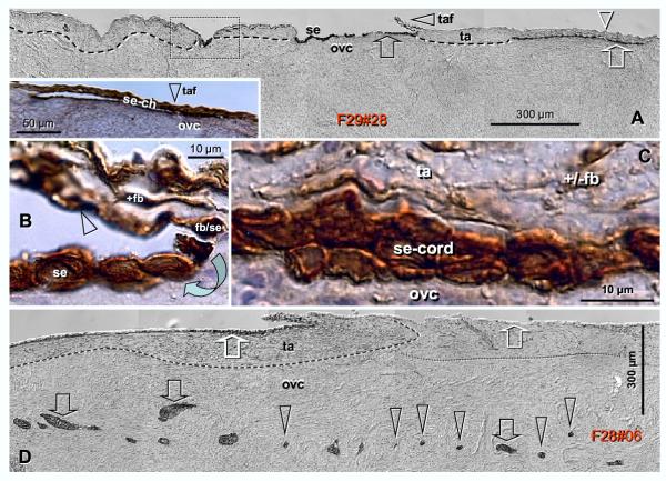
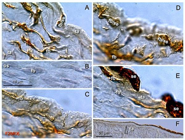
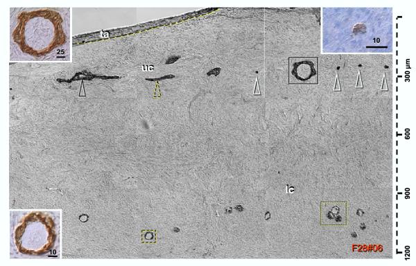
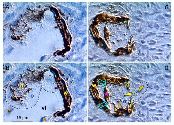
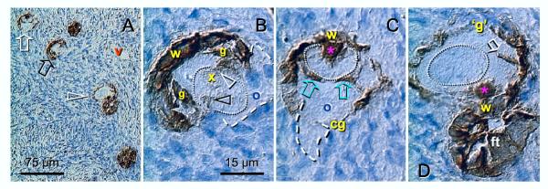
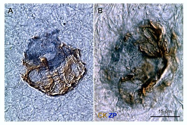
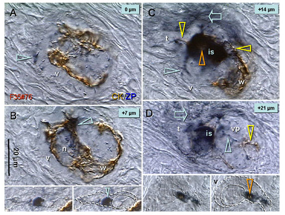
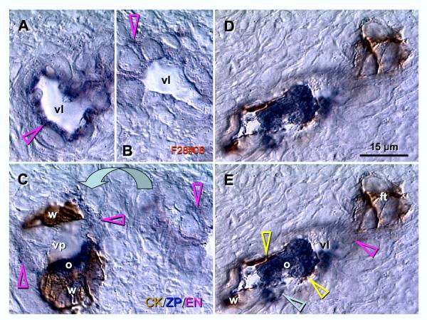
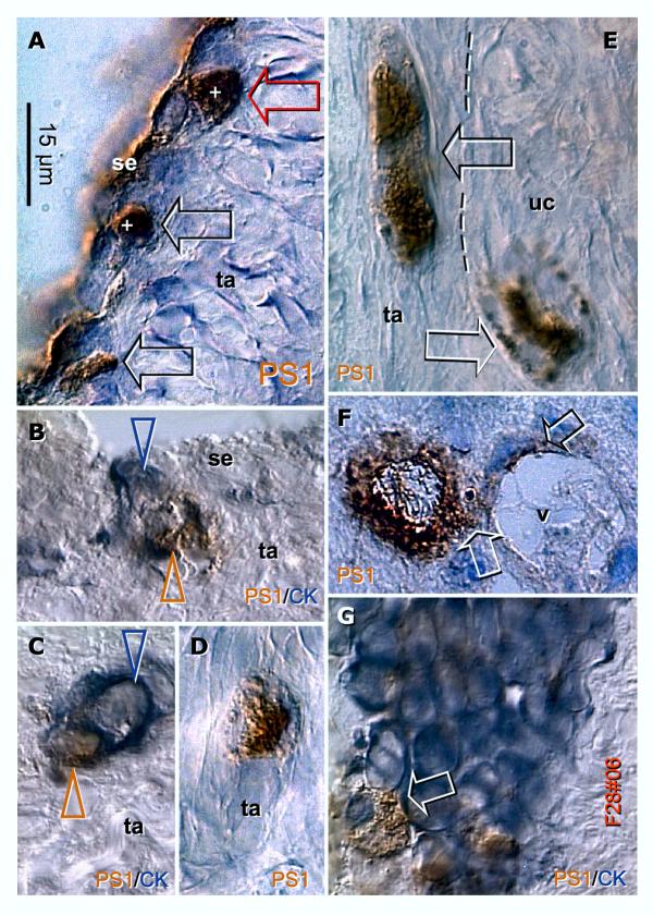
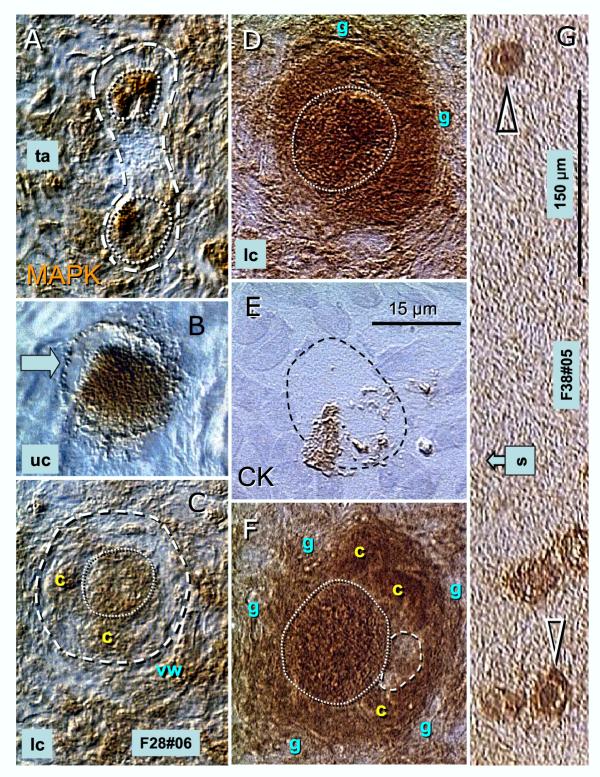
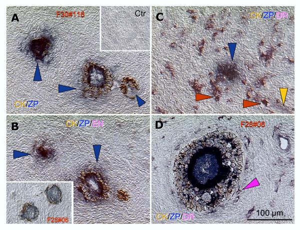
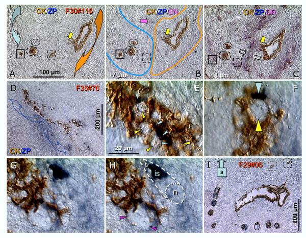
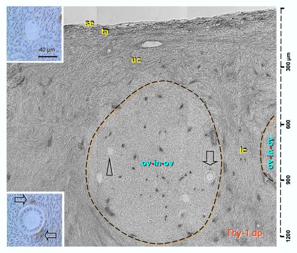
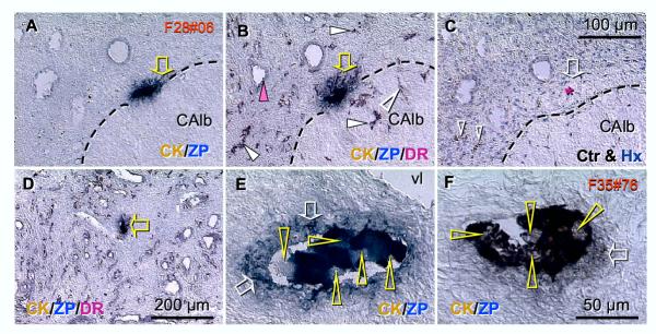
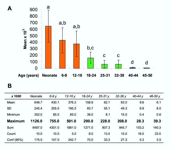
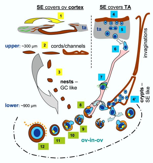
Recent reports indicate that functional mouse oocytes and sperm can be derived in vitro from somatic cell lines. We hypothesize that in adult human ovaries, mesenchymal cells in the tunica albuginea (TA) are bipotent progenitors with a commitment for both primitive granulosa and germ cells. We investigated ovaries of twelve adult women (mean age 32.8 +/- 4.1 SD, range 27-38 years) by single, double, and triple color immunohistochemistry. We show that cytokeratin (CK)+ mesenchymal cells in ovarian TA differentiate into surface epithelium (SE) cells by a mesenchymal-epithelial transition. Segments of SE directly associated with ovarian cortex are overgrown by TA, forming solid epithelial cords, which fragment into small (20 micron) epithelial nests descending into the lower ovarian cortex, before assembling with zona pellucida (ZP)+ oocytes. Germ cells can originate from SE cells which cover the TA. Small (10 micron) germ-like cells showing PS1 meiotically expressed oocyte carbohydrate protein are derived from SE cells via asymmetric division. They show nuclear MAPK immunoexpression, subsequently divide symmetrically, and enter adjacent cortical vessels. During vascular transport, the putative germ cells increase to oocyte size, and are picked-up by epithelial nests associated with the vessels. During follicle formation, extensions of granulosa cells enter the oocyte cytoplasm, forming a single paranuclear CK+ Balbiani body supplying all the mitochondria of the oocyte. In the ovarian medulla, occasional vessels show an accumulation of ZP+ oocytes (25-30 microns) or their remnants, suggesting that some oocytes degenerate. In contrast to males, adult human female gonads do not preserve germline type stem cells. This study expands our previous observations on the formation of germ cells in adult human ovaries. Differentiation of primitive granulosa and germ cells from the bipotent mesenchymal cell precursors of TA in adult human ovaries represents a most sophisticated adaptive mechanism created during the evolution of female reproduction. Our data indicate that the pool of primary follicles in adult human ovaries does not represent a static but a dynamic population of differentiating and regressing structures. An essential mission of such follicular turnover might be elimination of spontaneous or environmentally induced genetic alterations of oocytes in resting primary follicles.


Figures

Figure 1
Figure 1
Formation of epithelial cords from ovarian TA mesenchymal precursors - see also Ref [32]. A) Panoramic view of ovarian surface and adjacent cortex. Dashed line indicates interface between TA and stroma of the ovarian cortex. Dotted box indicates an early appearance of SE cells adjacent to the ovarian cortical stroma. Inset, epithelial channel (se-ch); se and arrow, surface epithelium cells; taf and arrowhead, TA flap; white arrowhead – a lack of SE cells above the TA; white arrow -bilaminar epithelial cord. B) Detail from (A) shows association of CK+ (brown color) fibroblasts (+fb,) with the TA flap surface (arrowhead), and transition from mesenchymal to epithelial morphology (fb/se) and surface epithelium cells (se, arched arrow). C) Detail from (A) shows CK+ epithelial cord consisting of two layers of epithelial cells and lying between the ovarian cortex (ovc) and TA. Note diminution of CK immunoexpression in TA fibroblasts (+/-fb). D) Parallel (black arrows) and perpendicular (arrowheads) view of epithelial cords in the upper ovarian cortex (ovc). Dashed line indicates a segment of CK+ TA with flap and differentiating SE cover (left white arrow). Dotted line indicates CK- segment lacking SE cower (right white arrowhead). F29 indicates female age in years; #28 = case No. Bar in (C), for (B and C). Cytokeratin 18 immunostaining (brown color) and hematoxylin counterstain.
Figure 2
Figure 2
Formation of ovarian SE from TA mesenchymal precursors. A) Tunica albuginea (ta) fibroblasts (fb) showing strong CK immunoexpression (brown color). One cell in mesenchymal-epithelial transition is apparent at the surface (fb/se). B) Ovarian stroma (os) shows no CK staining of fibroblasts (fb). C) Fibroblasts associated with the TA surface exhibit mesenchymal-epithelial transitory forms (D). E) Appearance of SE cells (se) is associated with a diminution of CK immunoexpression (-fb). F) Differentiation of the SE is associated with a diminution of cellularity in adjacent TA. Bar in (B), for (A-E). Cytokeratin 18 immunostaining, hematoxylin counterstain.
Figure 3
Figure 3
Distribution of epithelial cords and primary follicles in the ovarian cortex. Panoramic view composed of nine images shows ovarian tunica albuginea filled with CK18+ mesenchymal cells, upper cortex (uc) with epithelial channels (black arrowhead), cords (dashed and white arrowheads – see right inset) and follicle-like structures (solid box, see upper left inset for details). Lower cortex (Ic) shows isolated (dashed box – see lower left inset) and grouped primary follicles (dotted box). Bars in insets indicate μm. Cytokeratin 18 immunostaining, hematoxylin counterstain.
Figure 4
Figure 4
Association of epithelial nests with cortical vessels, oocyte-nest assembly and formation of CK18+ Balbiani body. A and B (copy of A with letters and dashed line) show the CK+ (brown color) nest body wall (w) inside of the cortical venule, which extends an arm (a) to catch the oocyte (o, dashed line) and move it outside of the blood vessel. C and D show the nest body and closing "gate" (g). A portion of the oocyte (dashed line) still lies outside of the complex, and is expected to move inside (arched arrow). The oocyte contains intraooplasmic CK+ (brown color) material (arrowheads). Extensions from the nest wall (arrows) contribute to the formation of CK+ paranuclear (Balbiani) body (asterisk). The oocyte nucleus is indicated by a dotted line. vl, vascular lumen; e, endothelial cells. CK18 immunostaining, hematoxylin counterstain.
Figure 5
Figure 5
Sequential stages of the oocyte-nest assembly and Balbiani body formation. A) A group of primary follicles in various stages of formation. Note adjacent venule (v). B) Early stage (see arrow in A) shows the oocyte during entrance into the CK+ (brown color) nest with nest wall (w) and inverted "gates" (g). Cytokeratin+ material appears to enter the nucleus (black arrowhead) and traces (white arrowhead) are visible around the nucleolus (x). Note a lack of Balbiani body. C) More advanced stage (white arrow in A) shows the oocyte nucleus (dotted line) adjacent to the nest wall (just opposite to the closing gate), embraced by intraooplasmic extensions (arrows). The Balbiani body (asterisk) appears to reside within the nucleus (dotted line), and small proportion of the ooplasm (dashed line) is still outside of the complex. D) Completed formation of primary follicle (arrowhead in A) shows closed gate ('g'), CK+ Balbiani body at the opposite side, adjacent to the nest wall, oocyte nucleus free of CK staining, and a single CK+ intraooplasmic extension (arrow) from the nest side. A follicle top (ft) from an adjacent follicle is also visible. CK18 immunostaining, hematoxylin counterstain.
Figure 6
Figure 6
Double color immunohistochemistry of the oocyte-nest assembly. A) Occupied "bird's" nest type indicates a half way oocyte-nest assembly. B) An earlier stage of assembly shows the oocyte embraced by nest trunks, resembling an octopus. Immunostaining with CK monoclonal antibody, clone MNF116 (recognizing CK5, 6, 8, 17) – first sequence visualized with diaminobenzidine (brown color), and with HSPZ (zona pellucida) polyclonal antibody – second sequence visualized with SG (blue color). No hematoxylin counterstain.
Figure 7
Figure 7
Serial sections through the intravascular oocyte-nest assembly. Serial sections (7 μm thick) through the oocyte-nest assembly show bottom of the complex (A); oocyte nucleus (n) plane (B); intermediate section (is) and the tail (t) plane (C); and top of the complex (D). v, vascular wall; vp, vascular pocket; blue arrowheads, ZP+ extensions from the oocyte intermediate section – anchors to the vascular wall; yellow arrowheads, nest extensions penetrating the ooplasm; blue arrows, ZP+ staining (oocyte signaling). Insets in panel B show putative germ cell with ZP+ intermediate section (arrowhead) migrating in the upper ovarian cortex. Insets in panel D show an association of putative germ cell with a cortical vessel (v) wall – note ZP+ intermediate section containing a CK+ "eye" (arrowhead). Details in text. Immunostaining with CK18 (brown color) and HSPZ (blue color). No hematoxylin counterstain.
Figure 8
Figure 8
Triple color immunohistochemistry for vascular route of the oocyte-nest assembly. Immunostaining CK/ZP/EN with CK18 (brown color), HSPZ (blue color), and endothelial CD31 – third sequence visualized with VIP(purple color). A) Medullary vessels show strong CD31 immunostaining (arrowhead), contrasting weak immunoexpression in cortical vessels (B). C) in the area adjacent to (B) (arched arrow) the vascular "pocket" (vp) shows adjacent endothelial cells, CK+ nest walls (w), and a ZP+ intermediate segment of the oocyte (o). Panels D and E show vascular wall (red arrowhead) with a lumen (vl) containing ZP+ oocyte "caught" by CK+ extensions (yellow arrowheads) from the nest wall (w). ft, follicle top of an adjacent primary follicle. No hematoxylin counterstain.
Figure 9
Figure 9
Association of PS1 meiotically expressed oocyte carbohydrate protein with asymmetric division and migration of putative germ cells. A) Segments of SE show cytoplasmic PS1 (brown color) expression (se). Dividing SE cells give rise to cells exhibiting nuclear PS1 immunostaining (+ nuclei, asymmetric division) and descending from the SE (arrows) into tunica albuginea (ta). This is particularly evident in the cell marked with a red arrow. B) Except asymmetrically divided SE cell – note CK+ (blue color and arrowhead) and PS1+ (brown color and arrowhead) daughter cells, no PS1 or CK immunoexpression is apparent in this SE segment or in the panel (C). D) In TA, the putative germ cells increase in size, but nuclear PS1 immunostaining persists. They show a symmetric division (arrow, E) and exhibit development of cytoplasmic PS1 immunoexpression when entering the upper (uc) ovarian cortex (white arrow). In the cortex, the cells show diminution of nuclear and increase of cytoplasmic PS1 staining (white arrow, panel F), particularly when attached to the cortical vessels (v). In such case, the PS1 immunoexpression appears to be extended toward endothelial cells (black arrow). In some instances, the asymmetric division giving rise to the putative PS1+ (brown color) germ cells could be observed at the periphery of CK+ (blue color) cortical epithelial crypts (arrow, panel G). Single (PS1) or double color immunohistochemistry (PS1/CK) as indicated, no hematoxylin counterstain.
Figure 10
Figure 10
Mitogen-activated protein kinase immunoexpression (brown color) in dividing and differentiating germ cells. Symmetrically dividing putative germ cells dashed line, panel A) in the TA (ta) showed strong nuclear MAPK immunostaining dotted lines. The cells showed an increase in size in the upper cortex (uc, panel B). Further increase in size of putative germ cells was detected during vascular transport, and accompanied by an appearance of focal cytoplasmic MAPK immunoexpression (c, panel C). D) During assembly with CK+epithelial nest (see brown color, serial section in panel E), the oocytes exhibited strong MAPK immunostaining of both the nuclear (dotted line) and cytoplasmic regions. In freshly formed primary follicles (F, see panel G for low power magnification of D and F), the MAPK heavily stained cytoplasmic clusters (c) were apparent, except an isolated paranuclear (Balbiani) body (dashed line). Strong nuclear staining (dotted line) persisted. MAPK (A-D, F and G) and CK single color immunohistochemistry (G), with hematoxylin counterstain. "s" arrow in (G) points to the distant ovarian surface.
Figure 11
Figure 11
Influx of macrophages during follicular atresia. Double color CK (brown color)/ZP (blue) immunohistochemistry revealed that cohorts of primary and secondary follicles in certain areas of the ovarian cortex showed degenerative changes, characterized in particular by fragmentation of the oocyte structure and dispersion of ZP+ staining among poorly defined layer of granulosa cells and adjacent stroma (arrowheads, panel A; see inset for control immunostaining). Inset in (B) shows normal primary follicles in another case. Triple color staining (CK/ZP/DR, panel C) revealed numerous large DR+ (purple color) macrophages (red arrowheads) invading the area from adjacent vessels (yellow arrowhead). In contrast, a healthy growing and preantral follicle (panel D) shows an association of sporadic small DR+ macrophages with the developing theca (red arrowhead). No hematoxylin counterstain.
Figure 12
Figure 12
Epithelial crypts – a source of germ cells for the alternative pathway of primary follicle formation. In ovaries showing atresia of follicular cohorts, association of epithelial nests with epithelial crypts has been observed. Panels (A-C) show CK+ (brown color) small epithelial nests, as evidenced from serial sections (compare the content of solid and dashed squares and oval areas on the left side). In panel (B), weakly CD31+ (purple) adjacent vessel is indicated by a red arrow, solid white arrows in A-C indicate an extension from the crypt toward the nests, and open arrows in (C) – see also lines in (B), indicate interface between stromal sprouts carrying epithelial nests and the crypt. Panel (C) also shows an accumulation of DR+(purple) macrophages around the stromal sprouts, and at their interface in particular. D) Remnant of an epithelial crypt (red arrow) from another case shows migration of ZP+ (HSPZ, blue) particles and stromal staining from one side (arched arrow). E-H) Details from panel D show an appearance of single (yellow arrowheads) and dividing germ-like cells (red arrowheads) among regressing CK+ cells (brown color). Note ZP+ segments (blue color and arrowheads) associated with unstained round cells. Dashed line in (H) indicates tadpole-like germ cell with leading nucleus (dotted line), ZP+ (blue color) intermediate section (is), and unstained tail (t). Panel (I) shows an association of primary follicles with the cortical epithelial crypt. Dashed boxes indicate unassembled epithelial nests. Details in text. No hematoxylin counterstain.
Figure 13
Figure 13
Distribution of Thy-1 differentiation protein in the ovarian cortex – an "ovary within the ovary" pattern. Thy-1 dp was strongly expressed by TA fibroblasts (ta), and moderately in the upper (uc) and lower ovarian cortex (Ic) except areas showing an "ovary within the ovary" pattern (ov-in-ov) with virtually no Thy-1 dp immunoexpression except vascular pericytes and smooth muscle cells. These areas characteristically contained primary follicles (arrowhead and upper inset) some of which showed an increase in size accompanied by Thy-1 dp+ pericytes (arrow and lower inset). Hematoxylin counterstain, details in text.
Figure 14
Figure 14
Accumulation of oocytes and their remnants in some medullary vessels. Some medullary vessels showed an embolus-like accumulation of ZP+ (HSPZ, blue color) material, either at the structures showing vascular regression, e.g., corpus albicans (CAlb, panels A-C), or without apparent vascular regression (panel D). In some instances, a higher magnification revealed the presence of multiple unstained oocyte nuclei (arrowheads, panels E and F). No hematoxylin counterstain except control staining in panel (C). vl, adjacent vascular lumen. Further details in text.
Figure 15
Figure 15
Age-related changes in primary follicle numbers in both human ovaries (A) and descriptive statistics (B). A) Statistical analysis of transformed cumulative data [Y = Log(Y)] reported by Block [53,63,64] and Gougeon [54] shows P < 0.0001 for one-way ANOVA. Tukey-Kramer Multiple Comparison post-test revealed that compared to neonatal ovaries a significant difference in follicle number first appears in the age group of 18 to 24 years. However, no significant difference was observed during the 20 years of optimal reproductive period, between females 18–38 years of age. Yet, the ovaries obtained from 40–50 year old females showed a significantly lower number of primary follicles vs. all other age groups. B) Descriptive statistics indicates plateau of the maximum follicle numbers (highlighted) during the 6–16, 18–38, and 40–50 age periods, associated with a lack of statistical significance between mean values ± SD – see panel (A).
Figure 16
Figure 16
Working model of possible pathways for formation of primary follicles in adult human ovaries (updated from Ref. [32]). 1) Ovarian tunica albuginea (ta) stem cells (green color) differentiate into the CK+ fibroblasts (red color) and by mesenchymal-epithelial transition give rise to the SE cells directly covering the ovarian cortex (arched arrow). 2) Closing of TA flaps (see Fig. 1) results in the formation of epithelial cords/channels in the upper ovarian cortex (see Fig. 3). Fragmented epithelial cords give rise to the epithelial nests, which resemble primitive granulosa cells [22] and descend into the lower ovarian cortex. 4) Depending on certain in situ (stromal) influences, the TA progenitors differentiate into the SE cells covering TA, which may, by asymmetric division, give rise to the ZP+ germ cells. 5) These putative germ cells may symmetrically divide, descend into the ovarian cortex, and associate with adjacent cortical vessels (6). Intravascular transport (7) is associated with a substantial increase of germ cell size and with development of ZP+ anchors (green lines), which may serve to slow down the transport speed and signal the epithelial nests to associate with a particular vascular segment. 8) The intravascular germ cells differentiating into the oocytes are picked up by epithelial nests associated with the proper cortical vessels. Such oocyte-nest complexes show an "octopus-like" (9) formations during the early stage of assembly, and a formation of the Balbiani body during the intermediate stage (yellow body, 10). The Balbiani body persists in resting primary follicles (11), but diminishes upon the growth promoting signals, including Thy-1 dp signaling derived from the follicle-accompanying vessels (12, dashed line). An alternative pathway for the germ cell origin from TA precursors (4') consists of a constitution of cortical crypts formed by SE-like embryonal type cells [22,25], possibly originating from, but not necessarily connected with, the deep SE invaginations, as evidenced from serial sections. The "alternative" pathway of germ cell origin may supply the oocytes directly to the neighboring nests (dashed arched arrows) and, via vascular transport (dotted arched arrow), saturate distant nests to form the primary follicles.

Comment in

References

    1. Waldeyer W. Eierstock und Ei Leipzig: Engelmann. 1870.
    1. Weissmann A. Die Continuitat des Keimplasmasals Grundlage einer Theorie der Vererbung. Jena: Fischer-Verlag; 1885.
    1. Franchi LL, Mandl AM, Zuckerman S. The development of the ovary and the process of oogenesis. In: Zuckerman S, editor. In The Ovary. London: Academic Press; 1962. pp. 1–88.
    1. Baker TG. Oogenesis and ovarian development. In: Balin H, Glasser S, editor. In Reproductive Biology. Amsterdam: Excerpta Medica; 1972. pp. 398–437.
    1. McLaren A. Signaling for germ cells. Genes Dev. 1999;13:373–376. - PubMed

MeSH terms

Substances