. 2004 Apr 28;32(8):2378-85.
doi: 10.1093/nar/gkh559. Print 2004.

Meiosis-specific yeast Hop1 protein promotes synapsis of double-stranded DNA helices via the formation of guanine quartets

Affiliations

Meiosis-specific yeast Hop1 protein promotes synapsis of double-stranded DNA helices via the formation of guanine quartets

S Anuradha et al. Nucleic Acids Res. .

Abstract

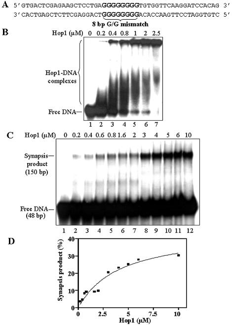
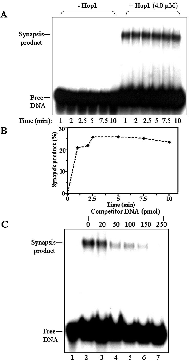
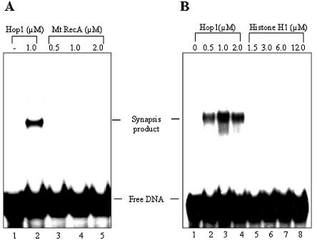
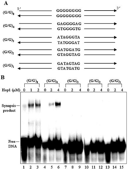
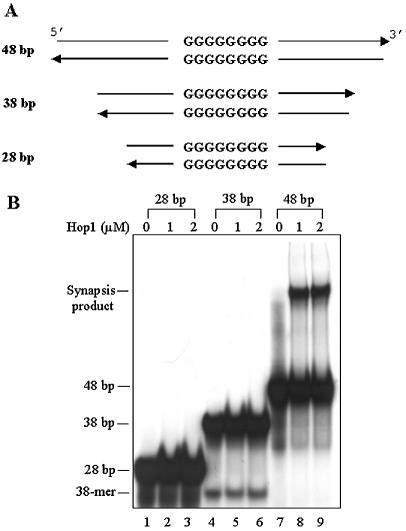
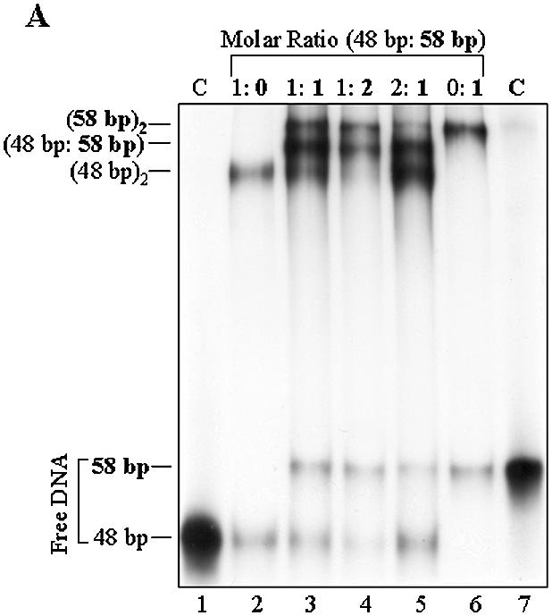
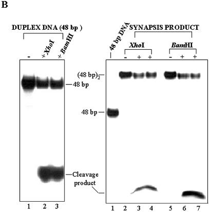
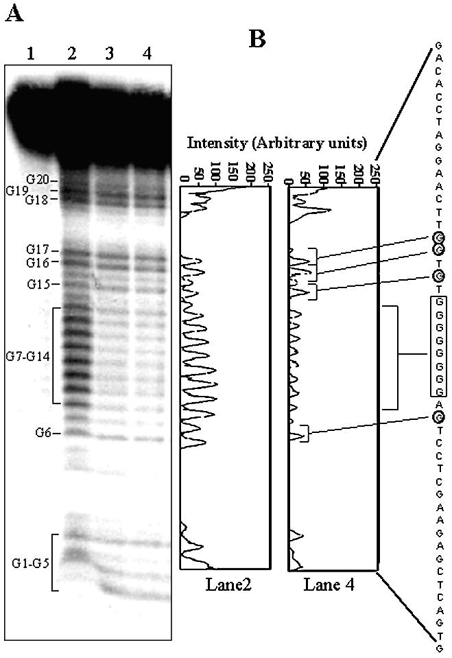
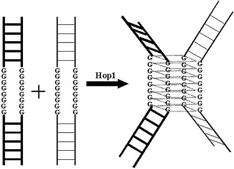
In most eukaryotes, genetic exchange between paired homologs occurs in the context of a tripartite proteinaceous structure called the synaptonemal complex (SC). Genetic analyses have revealed that the genes encoding SC proteins are vital for meiotic chromosome pairing and recombination. However, the number, nature and/or the mechanism used by SC proteins to align chromosomes are yet to be clearly defined. Here, we show that Saccharomyces cerevisiae Hop1, a component of SC, was able to promote pairing of double-stranded DNA helices containing arrays of mismatched G/G sequences. Significantly, pairing was rapid and robust, independent of homology in the arms flanking the central G/G region, and required four contiguous guanine residues. Furthermore, data from truncated DNA double helices showed that 20 bp on either side of the 8 bp mismatched G/G region was essential for efficient synapsis. Methylation interference indicated that pairing between the two DNA double helices involves G quartets. These results suggest that Hop1 is likely to play a direct role in meiotic chromosome pairing and recombination by its ability to promote synapsis between double-stranded DNA helices containing arrays of G residues. To our knowledge, Hop1 is the first protein shown to promote synapsis of DNA double helices from yeast or any other organism.


Figures

Figure 1
Figure 1
Hop1 binds to and promotes synapsis between double-stranded DNA containing mismatched G/G sequences. (A) Schematic of the DNA substrate. (B) Binding of Hop1 to 48 bp duplex DNA containing a mismatched G/G region. Reactions were performed with 0.5 µM of 32P-labeled 48 bp duplex DNA containing 8 bp mismatched G/G sequence in the absence or presence of indicated amounts of Hop1. (C) Hop1 promotes interstitial synapsis between double-stranded DNA helices containing 8 bp mismatched G/G sequence. Samples were deproteinized and analyzed as described in Materials and Methods. (D) Quantification of Hop1-promoted synapsis. The autoradiogram shown in (C) was scanned and the data were plotted as described in Materials and Methods.
Figure 2
Figure 2
Specificity of synapsis promoted by Hop1. (A) Kinetics of synapsis. 32P-labeled 48 bp duplex DNA containing an 8 bp G/G array (0.5 µM) was incubated in the absence (lanes 1–6) or presence (lanes 7–12) of Hop1 (4 µM) for varying time periods. Samples were deproteinized and analyzed as described in Materials and Methods. (B) Quantification of the kinetics of Hop1-promoted synapsis of DNA double helices. (C) Competitive inhibition of Hop1-promoted synapsis. Reaction mixtures contained 5 pmol of 32P-labeled 48 bp duplex DNA harboring an 8 bp G/G array, 4 µM Hop1 and an amount of unlabeled homologous duplex DNA as indicated above each lane.
Figure 3
Figure 3
RecA or histone H1 fail to promote synapsis of 48 bp duplex DNA containing an 8 bp G/G array. (A) Synapsis assay with Hop1 protein in comparison with MtRecA. 32P-labeled 48 bp duplex DNA containing an 8 bp G/G array (0.5 µM) was incubated in the absence (lane 1) or presence of Hop1 (lane 2) or MtRecA protein (lanes 3–5). Samples were deproteinized and analyzed as described in Materials and Methods. (B) Histone H1 fails to promote synapsis of 48 bp duplex DNA with a mismatched G/G array. Reactions were performed as described for (A). Lanes 1–8 represent reactions performed in the absence (lane 1) or presence of Hop1 (lanes 2–4) or histone H1 (lanes 5–8).
Figure 4
Figure 4
Mutations in the G/G region affect synapsis between double-stranded DNA helices. (A) Schematic of the DNA substrates. The mismatched G/G base pair is shown in bold face. (B) Hop1-promoted synapsis of DNA bearing a mutation in the G/G region. Each of the indicated 32P-labeled duplex DNAs (0.5 µM) was incubated with amounts of Hop1 as shown above each lane, at 30°C for 10 min. The reactions were deproteinized and analyzed as described in Materials and Methods. The subscript to the G/G parenthesis denotes the number of contiguous mismatched G/G base pairs.
Figure 5
Figure 5
Minimum sequence required for Hop1-promoted synapsis between two double-stranded DNA helices. (A) Schematic of the DNA substrates. (B) Synapsis between duplex DNA of varying length. Reaction mixtures contained 0.5 µM of 32P-labeled duplex DNA and Hop1p as indicated above each lane. Samples were incubated at 30°C for 10 min, deproteinized and analyzed as described in Materials and Methods. The position of duplex DNA and synapsis product are indicated on the left.
Figure 6
Figure 6
Hop1 promotes synapsis between heterologous double-stranded DNA helices. (A) Synapsis promoted by Hop1. Reaction mixtures containing Hop1 and 32P-labeled 48 bp and/or 58 bp DNA fragments were incubated at the specified molar ratios as indicated above each lane. Samples were deproteinized and analyzed as described in Materials and Methods. The positions of duplex DNAs and synapsis products are indicated on the left. (B) Synapsis between duplex DNA does not involve arms flanking the G/G region. Assays were performed in the presence or absence of specified restriction enzymes according to the manufacturer’s instructions. The cleavage products in lanes 3–4 and 6–7 have merged, resulting in a single band.
Figure 6
Figure 6
Hop1 promotes synapsis between heterologous double-stranded DNA helices. (A) Synapsis promoted by Hop1. Reaction mixtures containing Hop1 and 32P-labeled 48 bp and/or 58 bp DNA fragments were incubated at the specified molar ratios as indicated above each lane. Samples were deproteinized and analyzed as described in Materials and Methods. The positions of duplex DNAs and synapsis products are indicated on the left. (B) Synapsis between duplex DNA does not involve arms flanking the G/G region. Assays were performed in the presence or absence of specified restriction enzymes according to the manufacturer’s instructions. The cleavage products in lanes 3–4 and 6–7 have merged, resulting in a single band.
Figure 7
Figure 7
(A) Methylation interference implicates involvement of G residues in Hop1-promoted synapsis of DNA double helices. The assay was performed as described in Materials and Methods. In each lane, an equivalent amount of radioactivity was loaded to avoid differences in loading of the samples. Lane 1, reaction in which the duplex DNA was not treated with piperidine; lane 2, methylation pattern of the 48 bp duplex DNA; lanes 3 and 4, cleavage pattern of synapsis product generated in the presence of 2.5 and 5 µM Hop1, respectively. (B) The relative band intensity in lanes 2 and 4 in (A) was traced and plotted as arbitrary units. The protected guanines in the G/G region are boxed, and those in the flanking region are circled.
Figure 8
Figure 8
Schematic of synapsis promoted by Hop1. In the presence of Hop1, four G residues interact to form a G quartet by Hoogsteen base pairing. The proposed scheme is similar to the one proposed by Venczel and Sen (24). For details, see text.

References

    1. Roeder G.S. (1997) Meiotic chromosomes: it takes two to tango. Genes Dev., 11, 2600–2621. - PubMed
    1. Zickler D. and Kleckner,N. (1999) Meiotic chromosomes: integrating structure and function. Annu. Rev. Genet., 33, 603–754. - PubMed
    1. Sym M. and Roeder,G.S. (1994) Crossover interference is abolished in the absence of a synaptonemal complex protein. Cell, 79, 283–292. - PubMed
    1. Bishop D.K., Park,D., Xu,L. and Kleckner,N. (1992) DMC1: a meiosis-specific yeast homolog of E. coli recA required for recombination, synaptonemal complex formation and cell cycle progression. Cell, 69, 439–456. - PubMed
    1. Leu J.-Y. and Roeder,G.S. (1999) The pachytene checkpoint in S. cerevisiae depends on Swe1-mediated phosphorylation of the cyclin-dependent kinase Cdc28. Mol. Cell, 4, 805–814. - PubMed

Publication types

MeSH terms