. 2004 Apr 28;24(17):4250-8.
doi: 10.1523/JNEUROSCI.3920-03.2004.

Early striatal dendrite deficits followed by neuron loss with advanced age in the absence of anterograde cortical brain-derived neurotrophic factor

Affiliations

Early striatal dendrite deficits followed by neuron loss with advanced age in the absence of anterograde cortical brain-derived neurotrophic factor

Zachary C Baquet et al. J Neurosci. .

Abstract

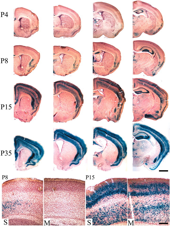
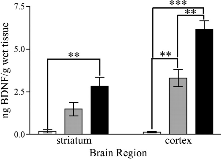
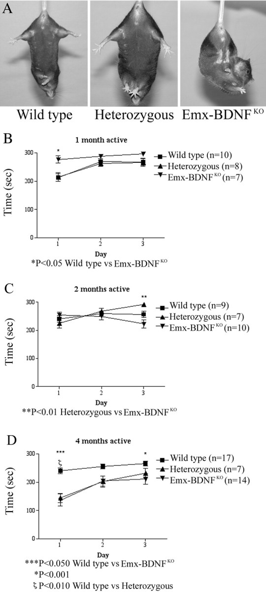
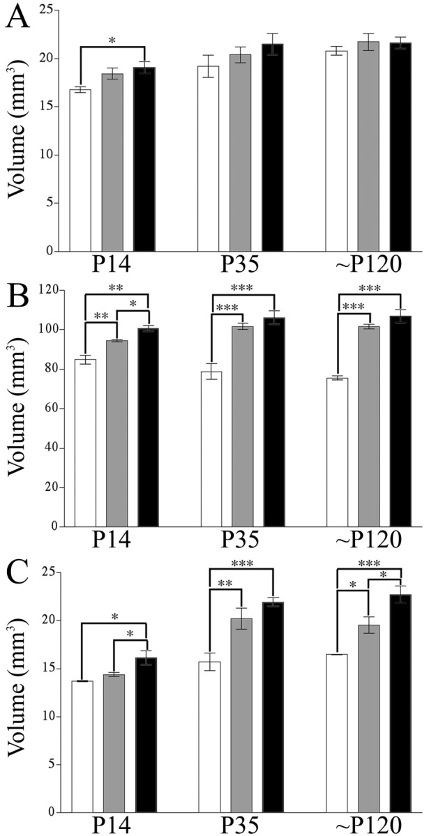
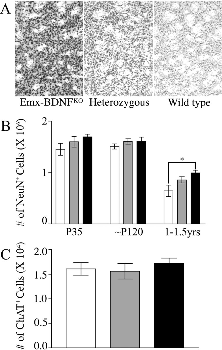
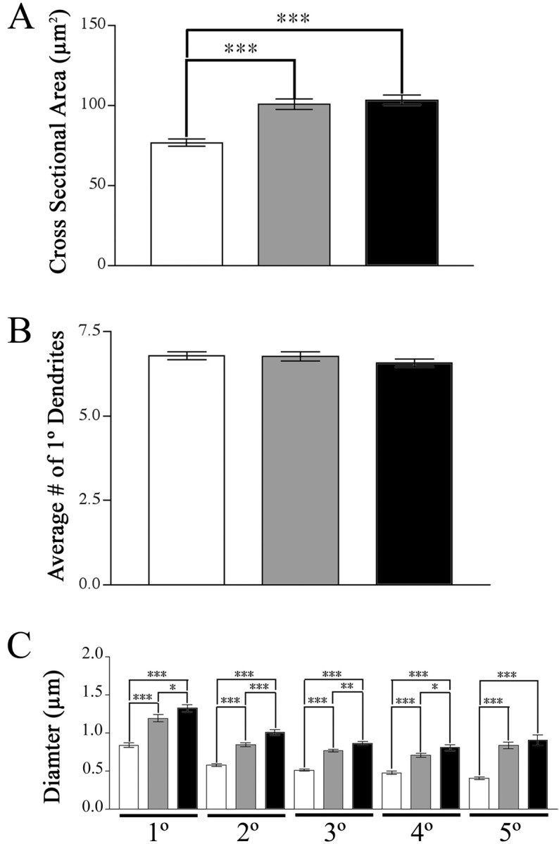
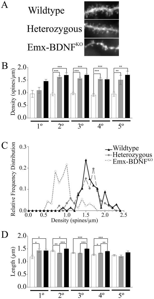
Brain-derived neurotrophic factor (BDNF), a member of the neurotrophin family, modulates neuronal survival, differentiation, and synaptic function. Reduced BDNF expression in the cortex caused by mutation of the huntingtin gene has been suggested to play a role in the striatal degeneration observed in Huntington's disease. BDNF expression rises dramatically in the cortex during the first few weeks of postnatal life in mice. Previously, it has been impossible to study the specific long-term effects of BDNF absence on CNS structures because of the early postnatal lethality of BDNF-/- mice. Mice harboring a floxed BDNF gene were bred with Emx1(IREScre/+) mice to generate Emx-BDNF(KO) mice that lack cortical BDNF but are viable. Adult Emx-BDNF(KO) mice display a hindlimb clasping phenotype similar to that observed in mouse models of Huntington's disease. The striatum of postnatal Emx-BDNF(KO) mice was reduced in volume compared with controls, and the most abundant neuron type of the striatum, medium spiny neurons (MSNs), had shrunken cell somas, thinner dendrites, and fewer dendritic spines at 35 d of age. Although significant striatal neuron losses were not detected at 35 or 120 d postnatal, 35% of striatal neurons were missing in Emx-BDNF(KO) mice aged beyond 1 year. Thus, cortical BDNF, although not required for the generation or near-term survival of MSN, is necessary for normal striatal neuron dendrite morphology during the period when BDNF expression rises in the cortex. Furthermore, a long-term in vivo requirement for cortical BDNF in supporting the survival of MSNs is revealed.


Figures

Figure 1.
Figure 1.
The striatum lacks apparent BDNF expression. BDNFlacZ/+ mice demonstrate substantial expression of the BDNF gene within the cortex with undetectable levels within the striatum. Shown are images (20×)of 40 μm coronal sections in anterior to posterior series from P4, P8, P15, and P35 mice stained with X-gal and counterstained with Neutral Red. Scale bar, 1 mm. At the bottom, 100× images of somatosensory (S) and motor (M) cortices of P8 and P15 mice. Scale bar, 200 μm.
Figure 2.
Figure 2.
Quantification of BDNF protein in cortex and striatum at P35. BDNF protein was quantified by ELISA for striatum and dorsal cortex and expressed as nanograms of BDNF protein per gram of wet tissue. These extracts were obtained from P35 mice; n = 3 mice per genotype (*p<0.05;**p<0.01;***p<0.001; one-way ANOVA with a Tukeypost hoctest). White bars are Emx-BDNFKO, gray bars are heterozygotes, and black bars are wild type.
Figure 3.
Figure 3.
Emx-BDNFKO mice exhibit a foot-clasping phenotype. Emx-BDNFKO, heterozygous, and wild-type mice of ages 1, 2, and 4 months were suspended by their tails for 1 min. Clasping was defined as the balling up of one or both of the hindlimb paws, accompanied by pulling them into the body and movement of the limbs toward the midline. A, Four-month-old wild-type, heterozygous, and Emx-BDNFKO mice undergoing tail suspension. B, Rotarod performance of 1-month-old mice (wild type, n = 10; heterozygotes, n = 8; Emx-BDNFKO, n = 7; *p < 0.05 for Emx-BDNFKO vs wild type). C, Two-month-old mice (wild type, n = 9; heterozygotes, n = 7; Emx-BDNFKO, n = 10; **p < 0.01 for heterozygous vs Emx-BDNFKO). D, Four-month-old mice (wild type, n = 17; heterozygotes, n = 7; Emx-BDNFKO, n = 14; *p < 0.05 wild type vs Emx-BDNFKO; ζp < 0.01 wild type vs heterozygous; ***p < 0.001 wild type vs Emx-BDNFKO). Squares are wild type, triangles are heterozygotes, and circles are Emx-BDNFKO.
Figure 4.
Figure 4.
CNS volume reduction in Emx-BDNFKO mice at P14, P35, and ∼P120. Specific CNS structures are reduced in volume in the absence of BDNF as assessed by the Cavalieri estimator. A, Hippocampus. B, Neocortex. C, Striatum (*p < 0.05; **p < 0.01; ***p < 0.001; one-way ANOVA with a Tukey post hoc test). White bars are Emx-BDNFKO, gray bars are heterozygotes, and black bars are wild type.
Figure 5.
Figure 5.
Loss of striatal neurons in old but not young adult Emx-BDNFKO mice. A, Representative images of striatum from all three genotypes immunostained for NeuN and counter-stained with cresyl violet (40 μm cryostat sections at 100× magnification). B, Optical fractionator estimate of the number of NeuN-positive cells in the striatum of P35, ∼P120, and 1- to 1.5-year-old mice. C, Estimate of the number of ChAT-positive cells in the striatum of ∼P120 mice. White bars are Emx-BDNFKO, gray bars are heterozygotes, and black bars are wild type.
Figure 6.
Figure 6.
Reduced soma and dendrite size inEmx-BDNFKO mice at P35. Neurons were labeled using DiOlistics and analyzed using fluorescent deconvolution microscopy. A, Cross-sectional areas of medium spiny neurons of the striatum (Emx-BDNFKO, 76.66 ± 2.14 μm2; heterozygous, 100.7 ± 3.32 μm2; wild type, 103.3 ± 3.08 μm2; represented as mean ± SEM). B, Number of primary dendrites on MSNs from each of the genotypes. C, Diameters of primary through fifth-order dendrites (*p < 0.05; **p < 0.01; ***p < 0.001; one-way ANOVA with a Tukeypost hoctest). White bars areEmx-BDNFKO, gray bars are heterozygotes, and black bars are wild type.
Figure 7.
Figure 7.
Reduced MSN spine density and length in Emx-BDNFKO mice. A, Representative images of DiI-labeled dendrite segments from the various genotypes at P35. B, Spine density per unit length of dendrite for different orders of dendrites. C, Relative frequency distributions of spine densities for each of the genotypes for pooled dendritic orders. D, Spine lengths for multiple orders of dendrites (Emx-BDNFKO as percentage of wild type: 1°, 90.1%, p < 0.05; 2°, 83.6%, p < 0.001; 3°, 89.1%, p < 0.01; 4°, 87.4, p < 0.05; 5°, 92.3%, p > 0.05; *p < 0.05; **p < 0.01; ***p < 0.001; one-way ANOVA with a Tukey post hoc test). White bars are Emx-BDNFKO, gray bars are heterozygotes, and black bars are wild type.

References

    1. Alberch J, Perez-Navarro E, Canals JM (2002) Neuroprotection by neurotrophins and GDNF family members in the excitotoxic model of Huntington's disease. Brain Res Bull 57: 817-822. - PubMed
    1. Alcantara S, Frisen J, del Rio JA, Soriano E, Barbacid M, Silos-Santiago I (1997) TrkB signaling is required for postnatal survival of CNS neurons and protects hippocampal and motor neurons from axotomy-induced cell death. J Neurosci 17: 3623-3633. - PMC - PubMed
    1. Altar CA, DiStefano PS (1998) Neurotrophin trafficking by anterograde transport. Trends Neurosci 21: 433-437. - PubMed
    1. Altar CA, Cai N, Bliven T, Juhasz M, Conner JM, Acheson AL, Lindsay RM, Wiegand SJ (1997) Anterograde transport of brain-derived neurotrophic factor and its role in the brain. Nature 389: 856-860. - PubMed
    1. Andrew SE, Goldberg YP, Kremer B, Telenius H, Theilmann J, Adam S, Starr E, Squitieri F, Lin B, Kalchman MA, et al. (1993) The relationship between trinucleotide (CAG) repeat length and clinical features of Huntington's disease. Nat Genet 4: 398-403. - PubMed

Publication types

MeSH terms

Substances