. 2004 May 19;23(10):2059-70.
doi: 10.1038/sj.emboj.7600159. Epub 2004 Apr 29.

Entry of newly synthesized GLUT4 into the insulin-responsive storage compartment is GGA dependent

Affiliations

Entry of newly synthesized GLUT4 into the insulin-responsive storage compartment is GGA dependent

Robert T Watson et al. EMBO J. .

Abstract

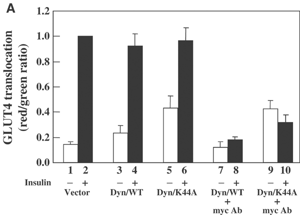
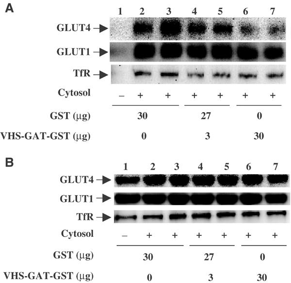
Following biosynthesis, both GLUT1 and VSV-G proteins appear rapidly (2-3 h) at the plasma membrane, whereas GLUT4 is retained in intracellular membrane compartments and does not display any significant insulin responsiveness until 6-9 h. Surprisingly, the acquisition of insulin responsiveness did not require plasma membrane endocytosis, as expression of a dominant-interfering dynamin mutant (Dyn/K44A) had no effect on the insulin-stimulated GLUT4 translocation. Furthermore, expression of endocytosis-defective GLUT4 mutants or continuous surface labeling with an exofacial specific antibody demonstrated that GLUT4 did not transit the cell surface prior to the acquisition of insulin responsiveness. The expression of a dominant-interfering GGA mutant (VHS-GAT) had no effect on the trafficking of newly synthesized GLUT1 or VSV-G protein to the plasma membrane, but completely blocked the insulin-stimulated translocation of newly synthesized GLUT4. Furthermore, in vitro budding of GLUT4 vesicles but not GLUT1 or the transferrin receptor was inhibited by VHS-GAT. Together, these data demonstrate that following biosynthesis, GLUT4 directly sorts and traffics to the insulin-responsive storage compartment through a specific GGA-sensitive process.


Figures

Figure 1
Figure 1
Newly synthesized VSV-G and GLUT1 proteins rapidly accumulate at the plasma membrane, whereas GLUT4 is retained in the perinuclear region. (A) Differentiated 3T3L1 adipocytes were electroporated with 50 μg of cDNAs encoding VSV-G (panels a–g), GLUT1-EGFP (panels h–n) or GLUT4-EGFP (panels o–u). Following transfection, the cells were immediately incubated for 2–12 h, fixed and processed for confocal fluorescent microscopy. The VSV-G protein was detected with a monoclonal VSV-G antibody followed by a Texas Red-conjugated secondary antibody. GLUT1-EGFP and GLUT4-EGFP were detected by GFP fluorescence. These are representative experiments independently performed 3–4 times. (B) Differentiated 3T3L1 adipocytes were electroporated with 50 μg GLUT4-EGFP cDNA and incubated for 3–12 h. The cells were then either left untreated (basal, panels a–f) or stimulated with 100 nm insulin (insulin, panels g–l) for 30 min. The cells were then fixed and processed for confocal fluorescent microscopy. These are representative experiments independently performed 3–5 times. (C) Differentiated 3T3L1 adipocytes were electroporated with 50 μg of cDNAs encoding GLUT1-EGFP (open symbols) or GLUT4-EGFP (closed symbols) and either untreated (circles) or incubated with 100 nm insulin for 30 min (squares) at the times indicated. The number of cells displaying a continuous EGFP rim fluorescence is expressed as the average from the counting of 100 cells from four independent experiments.
Figure 2
Figure 2
Electroporation does not disrupt insulin-stimulated translocation of the endogenous glut4 protein. (A) Differentiated 3T3L1 adipocytes were transfected with 50 μg of IRAP-EGFP cDNA and the cells incubated for 2.5 h. The cells were then either left untreated (basal, panels a–c) or incubated with 100 nm insulin (insulin, panels d–f) for 30 min. The samples were then fixed and examined for endogenous GLUT4 translocation using a GLUT4 polyclonal antibody (panels a and d) or GFP fluorescent for EGFP-IRAP translocation (panels b and e). The merged images are presented in panels c and f. This is a representative experiment independently performed three times. (B) 3T3L1 adipocytes were transfected with 50 μg of GLUT1-EGFP cDNA and the cells incubated for 2.5 h. The cells were then either left untreated (basal, panels a–c) or incubated with 100 nM insulin (insulin, panels d–f) for 30 min. Plasma membrane sheets were then prepared and labeled with a GLUT4 polyclonal antibody (panels a and d) or detected by GFP fluorescence for GLUT1-EGFP (panels b and e). The merged images are presented in panels c and f. This is a representative experiment independently performed four times.
Figure 3
Figure 3
Expression of a dominant-interfering dynamin mutant (Dyn/K44A) does not affect the time-dependent acquisition of insulin responsiveness. Differentiated 3T3L1 adipocytes were electroporated with 100 μg GLUT4-EGFP plus 100 μg HA-Dyn/WT (A) or HA-Dyn/K44A (B) cDNAs. Immediately following transfection, the cells were incubated with 5 μg/ml BFA for 3 h to allow dynamin expression but prevent GLUT4-EGFP exit from the endoplasmic reticulum. BFA was then removed and at the times indicated either untreated (basal, panels a–e) or treated with 100 nM insulin (insulin, panels f–j) for 30 min. Cells were labeled with an HA antibody and a Texas Red-conjugated donkey anti-mouse secondary antibody to detect dynamin expression. These are representative images obtained from 3–6 independent experiments. (C) The data were quantified by determining the number of adipocytes displaying a continuous GLUT4-EGFP rim fluorescence in cells coexpressing Dyn/WT (squares) and Dyn/K44A (circles) in the basal (open symbols) and insulin-stimulated (filled symbols) states. These data represent the average with standard deviation from the counting of 100 cells from four independent experiments.
Figure 4
Figure 4
The newly synthesized GLUT4 protein does not transit the plasma membrane prior to becoming insulin responsive. (A) Differentiated 3T3L1 adipocytes were electroporated with 100 μg myc-GLUT4-EGFP with and without 100 μg Dyn/WT or Dyn/K44A cDNAs. Immediately following transfection, the cells were left untreated (bars 1–6) or incubated with 2 μg/ml of the myc monoclonal antibody (bars 7–10) for 6 h at 37°C. The cells were then serum starved in the absence (bars 1–6) or presence of 2 μg/ml of the myc monoclonal antibody (bars 7–10) for 2.5 h. The cells were then either untreated (open bars) or treated with 100 nM insulin (filled bars) for 30 min. Thus, the total duration of time in these experiments was 9 h. The cells in bars 1–6 were washed, fixed, blocked and incubated with the primary myc antibody and then Texas Red-conjugated rabbit anti-mouse secondary antibody. The cells in bars 7–10 were washed to remove the unbound myc antibody, fixed, blocked and then directly incubated with the Texas Red-labeled rabbit anti-mouse secondary antibody. Quantification of myc-GLUT4-EGFP translocation was determined. These data represent the average with standard deviation from the quantification of 30 cells in three independent experiments.
Figure 5
Figure 5
Expression of an endocytosis-defective GLUT4 mutant does not impair insulin stimulation of the newly synthesized GLUT4 protein. Differentiated 3T3L1 adipocytes were electroporated with 50 μg of the wild-type GLUT4-EGFP fusion protein (A, GLUT4/WT), the carboxyl-terminal endocytosis-defective GLUT4-EGFP mutant (B, GLUT4/SAA) or the amino-terminal endocytosis-defective GLUT4-EGFP mutant (B, GLUT4/AQQI). At the times indicated, the cells were then either left untreated (filled circles) or stimulated with 100 nM insulin (open circles) for 30 min. The cells were then fixed and processed for confocal fluorescent microscopy. The data were quantified by determining the number of adipocytes displaying a continuous myc-GLUT4 rim fluorescence. These data represent the average with standard deviation from the counting of 100–150 cells from 4–6 independent experiments.
Figure 6
Figure 6
The acquisition of insulin-stimulated GLUT4 translocation is GGA dependent. (A) Differentiated 3T3L1 adipocytes were electroporated with 100 μg myc-GLUT4 plus 100 μg of the empty vector (pcDNA3). At various times following transfection, the cells were either left untreated (open circles) or stimulated with 100 nM insulin (filled circles) for 30 min. The cells were then fixed, labeled with a myc antibody and a Texas Red-conjugated donkey anti-mouse secondary antibody to detect myc-GLUT4 at the cell surface. The data were quantified by determining the number of adipocytes displaying continuous myc-GLUT4 rim fluorescence. These data represent the average with standard deviation from the counting of 100 cells from four independent experiments. (B) Differentiated 3T3L1 adipocytes were electroporated with 100 μg myc-GLUT4 plus 100 μg of the cDNA encoding the dominant-interfering GGA mutant EGFP fusion protein (VHS-GAT). The cells were either left untreated (open circles) or stimulated with 100 nM insulin (filled circles) and analyzed for GLUT4 translocation as described above. (C) Differentiated 3T3L1 adipocytes were electroporated with 100 μg of the cDNA encoding myc-GLUT1 plus 100 μg of the empty vector (filled squares) or 100 μg myc-GLUT1 cDNA plus 100 μg of the cDNA encoding the dominant-interfering GGA mutant EGFP fusion protein (open squares). At various times following transfection, the cells were fixed, labeled with a myc antibody and a Texas Red-conjugated donkey anti-mouse secondary antibody to detect myc-GLUT1 at the cell surface as described above. (D) Differentiated 3T3L1 adipocytes were electroporated with 100 μg of the cDNA encoding VSV-G protein plus 100 μg of the empty vector (filled squares) or 100 μg VSV-G cDNA plus 100 μg of the cDNA encoding the dominant-interfering GGA mutant EGFP fusion protein (open squares). These data represent the average with standard deviation from the counting of 100 cells from four independent experiments.
Figure 7
Figure 7
Insulin-stimulated GLUT4 trafficking from the GSC to the plasma membrane is independent of GGA function. (A) Differentiated 3T3L1 adipocyte nuclei were microinjected with 50 μg of the MBP-Ras fusion cDNA plus 200 μg of either the empty vector (panels a–c), VHS-GAT (panels d–f) or TC10 (panels g–i) cDNAs. The cells were allowed to recover for 24 h and either left untreated (panels a–c) or stimulated with 100 nM insulin (panels d–i) for 30 min. Plasma membrane sheets were then prepared and labeled with an MBP antibody (panels a, d, g) or a GLUT4 antibody (panels b, e, h). The merge images are shown in panels c, f and i. These are representative images obtained from three independent experiments with the visualization of 60 individual MBP-Ras-positive plasma membrane sheets. (B) These data were quantified by counting the number of plasma membrane sheets from microinjected cells that displayed endogenous GLUT4 translocation.
Figure 8
Figure 8
The dominant-interfering GGA mutant inhibits GLUT4 vesicle budding in vitro. (A) Golgi- and endosome-enriched donor membranes were incubated with ATP and GTPγS in the absence (lane 1) or presence (lanes 2–7) of cytosol and as described under Materials and methods. The reactions were also performed in the presence of 30 μg GST (lanes 1–3), 3 μg (lanes 4 and 5) and 30 μg (lanes 6 and 7) of the VHS-GAT-GST fusion protein. The released membrane vesicles (A) and Golgi imput membranes (B) were then subject to immunoblotting using GLUT4, GLUT1 and transferrin receptor (TfR) antibodies. This is a representative immunoblot independently performed four times.
Figure 9
Figure 9
Schematic representation of the trafficking of newly synthesized GLUT4 protein in adipocytes. GLUT4 protein undergoes co-translational insertion into the endoplasmic reticulum and traffics through the Golgi into the TGN. GLUT4 exit from the TGN is distinct from GLUT1 protein in that GLUT4 does not initially traffic to the plasma membrane but is targeted directly to the GSC. The exit of GLUT1 from the TGN is GGA independent, whereas the transport of GLUT4 from the TGN to the GSC is GGA dependent. In the basal state, GLUT4 slowly leaves the GSC en route to the plasma membrane where it is recycled back to the GSC through endocytosis and endosome sorting. In the presence of insulin, the transport of GLUT4 out of the GSC is markedly increased and exit from this compartment is GGA independent.

References

    1. Al-Hasani H, Hinck CS, Cushman SW (1998) Endocytosis of the glucose transporter GLUT4 is mediated by the GTPase dynamin. J Biol Chem 273: 17504–17510 - PubMed
    1. Baumann CA, Ribon V, Kanzaki K, Thurmond DC, Mora S, Shigematsu S, Bickel PE, Pessin JE, Saltiel AR (2000) CAP defines a second signaling pathway required for insulin-stimulated glucose transport. Nature 407: 202–207 - PubMed
    1. Bergmann JE (1989) Using temperature-sensitive mutants of VSV to study membrane protein biogenesis. Methods Cell Biol 32: 85–110 - PubMed
    1. Bogan JS, Lodish HF (1999) Two compartments for insulin-stimulated exocytosis in 3T3-L1 adipocytes defined by endogenous ACRP30 and GLUT4. J Cell Biol 146: 609–620 - PMC - PubMed
    1. Bryant NJ, Govers R, James DE (2002) Regulated transport of the glucose transporter GLUT4. Nat Rev Mol Cell Biol 3: 267–277 - PubMed

Publication types

MeSH terms