. 2004 Apr 26;165(2):191-202.
doi: 10.1083/jcb.200310139.

Poly(A)+ RNAs roam the cell nucleus and pass through speckle domains in transcriptionally active and inactive cells

Affiliations

Poly(A)+ RNAs roam the cell nucleus and pass through speckle domains in transcriptionally active and inactive cells

Chris Molenaar et al. J Cell Biol. .

Abstract

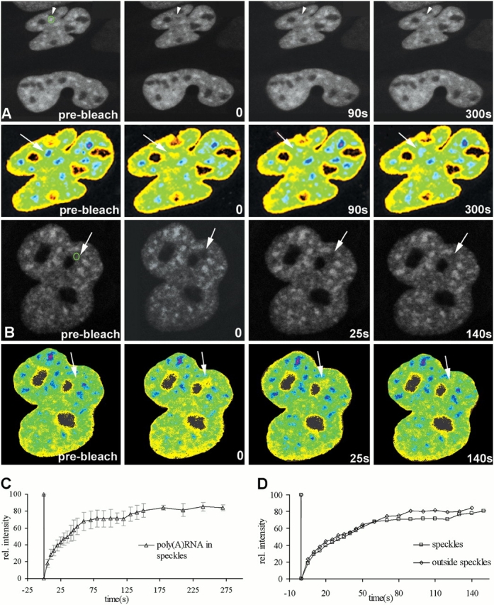
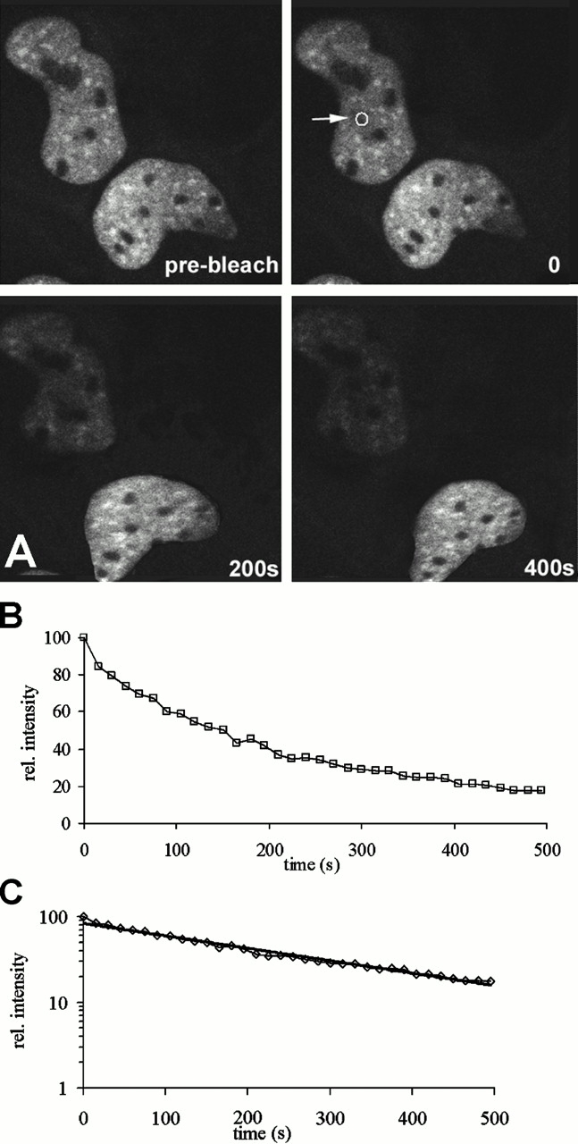
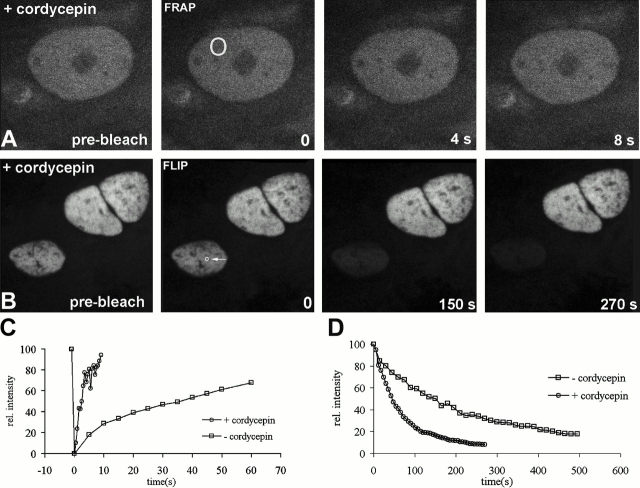
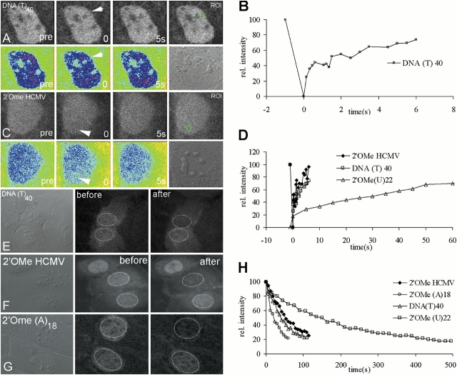
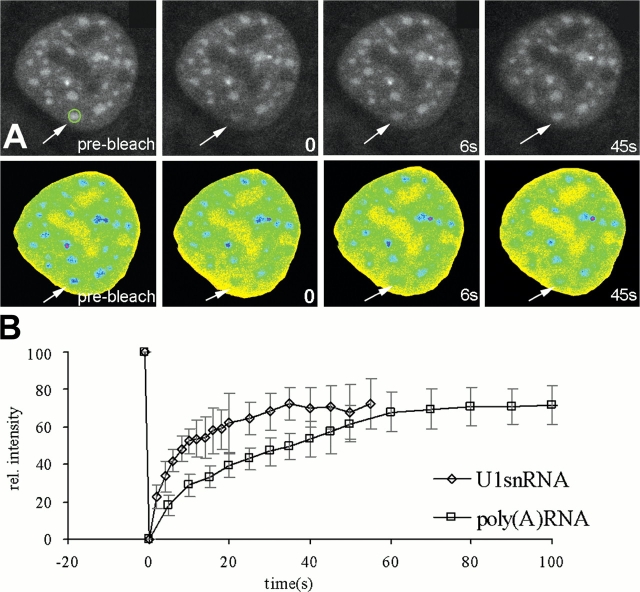
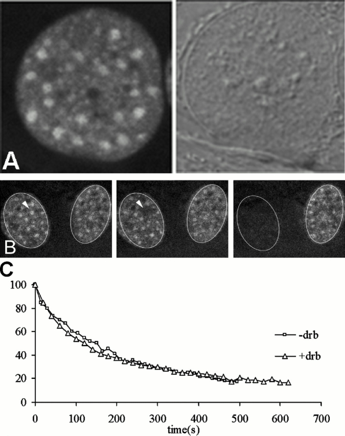
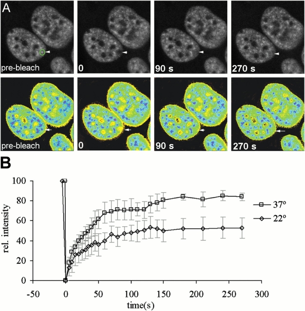
Many of the protein factors that play a role in nuclear export of mRNAs have been identified, but still little is known about how mRNAs are transported through the cell nucleus and which nuclear compartments are involved in mRNA transport. Using fluorescent 2'O-methyl oligoribonucleotide probes, we investigated the mobility of poly(A)+ RNA in the nucleoplasm and in nuclear speckles of U2OS cells. Quantitative analysis of diffusion using photobleaching techniques revealed that the majority of poly(A)+ RNA move throughout the nucleus, including in and out of speckles (also called SC-35 domains), which are enriched for splicing factors. Interestingly, in the presence of the transcription inhibitor 5,6-dichloro-1-beta-D-ribofuranosylbenzimidazole, the association of poly(A)+ RNA with speckles remained dynamic. Our results show that RNA movement is energy dependent and that the proportion of nuclear poly(A)+ RNA that resides in speckles is a dynamic population that transiently interacts with speckles independent of the transcriptional status of the cell. Rather than the poly(A)+ RNA within speckles serving a stable structural role, our findings support the suggestion of a more active role of these regions in nuclear RNA metabolism and/or transport.


Figures

Figure 1.
Figure 1.
FRAP analysis of fluorescent 2'OMe (U) 22 demonstrates that poly(A) + RNA is a dynamic component of speckles and nucleoplasm. Living U2OS cells injected with (U)22-TAMRA were subjected to FRAP analysis. A speckle (A) and a nucleoplasmic area outside a speckle (B) were selected and photobleached. Images were recorded just before bleaching and at different time intervals after bleaching. The green circles indicate the photobleached region. To illustrate the recovery of fluorescence more clearly, pseudocolor images of the bleached cells are shown. Fluorescence intensities range from yellow (low) to blue (high). The fluorescence recovery in the bleached areas, indicated by arrows and arrowheads, was quantified, and relative fluorescence intensities, which were calculated as described in Materials and methods, are displayed in recovery curves (C and D). The measurement immediately after bleaching was set at 0 s. In speckles, fluorescence recovery reached a plateau at ∼85% around 250 s (C). The error bars represent SD. (D) The recovery curve from a bleached area outside a speckle is compared with one obtained from a bleached speckle. The curves shown in C and D represent the average values of 15 measured cells.
Figure 2.
Figure 2.
FLIP analysis of fluorescent 2'OMe (U) 22 shows that poly(A) + RNA moves throughout the nucleus. (A) The fluorescence intensity of the top cell gradually decreases when a spot (arrow) is repeatedly bleached. (B) The corresponding FLIP curve shows that a relative immobile fraction of ∼15% remains present. (C) Semi-logarithmic plot of nuclear loss of fluorescence shows that the curve can be fitted to a single exponential fit, indicating a single population of slow moving poly(A)+ RNA.
Figure 3.
Figure 3.
Diffuse nuclear staining and high mobility rate of 2'OMe (U) 22 in cordycepin-treated cells. The localization pattern of 2'OMe (U)22 in cordycepin-treated cells reveals that the probe is diffusely spread throughout the nucleoplasm and not concentrated in speckles. Both FRAP (A and C) and FLIP (B and D) analysis revealed a mobility rate that is significantly higher compared with untreated cells, indicating that the mobility rate is strongly dependent on specific hybridization to poly(A)+ RNA. The circles indicate the bleached areas.
Figure 4.
Figure 4.
FRAP and FLIP analysis show that control probes are highly mobile. (A) In U2OS cells, the TAMRA-labeled (dT)40 probe reveals a diffuse staining of the nucleoplasm, excluding nucleoli, after cytoplasmic microinjection. Bleaching of a selected area (green circle and arrows) resulted in a rapid recovery of fluorescence. Due to the rapid movement of the probe, the fluorescence intensity immediately after bleaching had already returned to ∼80% of the initial fluorescence, whereas within a few seconds a recovery to ∼100% was observed. Quantitative analysis of the FRAP measurements confirms the high mobility of the (dT)40 probe (B) and shows that (dT)40 moves much faster through the nucleus compared with the 2'OMe (U)22 probe (D). Similar kinetics were observed using the control probe 2'OMe HCMV that has no target in U2OS cells (C and D). (C) The circle and arrowheads indicate the bleached areas. Fluorescence intensities are also represented in pseudocolor, and the differential interference contrast images are recorded after the bleaching experiment showing that the bleached cells are not affected by the experimental conditions. FLIP analysis of cells injected with (dT)40 (E), 2'OMe HCMV (F), and 2'OMe (A)18 (G) reveals that these probes move significantly faster through the nucleoplasm than the 2'OMe (U)22 probe (H).
Figure 5.
Figure 5.
2'OMe U1snRNA-TAMRA localizes to speckles and has a higher mobility rate than 2'OMe (U) 22. After cytoplasmic microinjection, 2'OMe U1 snRNA-TAMRA localizes to nucleoplasm, speckles, and Cajal bodies (two bright dots), but not to nucleoli. The mobility of this probe was analyzed by FRAP. Images were recorded just before, immediately after, and at regular time intervals after photobleaching. (A) 4 images out of a series of 26 are displayed, and the arrow indicates the speckle that has been photobleached. (B) The corresponding FRAP curve is plotted together with the FRAP curve for 2'OMe (U)22-TAMRA, indicating that U1snRNA is more dynamic than poly(A)+ RNA in cell nuclei. The error bars represent SD.
Figure 6.
Figure 6.
DRB treatment does not reduce the mobility of poly(A) + RNA. After microinjection of the 2'OMe (U)22-TAMRA probe, DRB was added to the medium to a final concentration of 50 μg/ml, and FLIP analysis was performed 3–4 h later. Incubation with DRB resulted in enlargement and rounding-up of speckles (A), whereas the morphology of the cell nucleus was not affected as shown in the differential interference contrast image. FLIP analysis of DRB-treated cells revealed no significant difference in the rate of fluorescence loss between treated and nontreated cells (B and C). The arrowhead is indicating the speckle that has been repeatedly bleached at high laser power.
Figure 7.
Figure 7.
The import of poly(A) + RNA into speckles is reduced at 22°C. Cells microinjected with 2'OMe (U)22-TAMRA were incubated at either 37or 22°C and subjected to FRAP analysis. At each temperature, 15 cells were analyzed. (A) Confocal images out of a series of 26 show the recovery of fluorescence within a speckle (green circle) at different time points after photobleaching (arrowheads and arrows) in cells kept at 22°C. (B) The recovery curves of cells incubated at 37 or 22°C are plotted with error bars representing SD.
Figure 8.
Figure 8.
SF2/ASF, PABP2, and Aly move more rapidly toward speckles than poly(A) + RNA. Cells expressing SF2/ASF-GFP were microinjected with 2'OMe (U)22-TAMRA and subjected to FRAP analysis (A). SF2/ASF-GFP and 2'OMe (U)22-TAMRA were simultaneously bleached in a speckle (arrowheads), and images were recorded before, just after, and at regular time intervals after bleaching. The images taken at 12 s after bleaching indicate that SF2/ASF-GFP (green) has a higher recovery rate compared with poly(A)+ RNA (red). In the combined image at 12 s, the green fluorescence is much stronger than the red. This difference in recovery rate is more evidently shown in the mask images in which areas where both SF2/ASF-GFP and poly(A)+ RNA are present above a threshold value are shown in white. Only after 120 s, a full recovery of fluorescence is observed. (B) FRAP was also performed on cells expressing PABP2-GFP. A speckle was selected (arrow) and photobleached, and the recovery of fluorescence was monitored. As shown, a full recovery of PABP2-GFP fluorescence was obtained within 50 s. Quantitative analysis of recoveries for SF2/ASF-GFP (n = 8) or PABP2-GFP (n = 12) show similar patterns for both proteins, reaching a plateau at ∼90% of the initial fluorescence after ∼40 s (D). For reasons of comparison, the recovery plot of poly(A)+ RNA is also shown. Cells expressing Aly-GFP were imaged before and after photobleaching a speckle in the nucleus (C, arrow). The images and the corresponding recovery curve (E) show that fluorescence recovered within 1 min. The error bars in D and E represent SD. The FLIP curve of Aly-GFP shows a complete loss of fluorescence within 80 s, which is a little slower than TAP-GFP (F).

Similar articles

Cited by

References

    1. Bauren, G., and L. Wieslander. 1994. Splicing of Balbiani ring 1 gene pre-mRNA occurs simultaneously with transcription. Cell. 76:183–192. - PubMed
    1. Bauren, G., W.Q. Jiang, K. Bernholm, F. Gu, and L. Wieslander. 1996. Demonstration of a dynamic, transcription-dependent organization of pre-mRNA splicing factors in polytene nuclei. J. Cell Biol. 133:929–941. - PMC - PubMed
    1. Braun, I.C., A. Herold, M. Rode, E. Conti, and E. Izaurralde. 2001. Overexpression of TAP/p15 heterodimers bypasses nuclear retention and stimulates nuclear mRNA export. J. Biol. Chem. 276:20536–20543. - PubMed
    1. Bridge, E., K.U. Riedel, B.M. Johansson, and U. Petterson. 1996. Spliced exons of adenovirus late RNAs colocalize with snRNP in a specific nuclear domain. J. Cell Biol. 135:303–314. - PMC - PubMed
    1. Calado, C., and M. Carmo-Fonseca. 2000. Localization of poly(A)-binding protein 2 (PABP2) in nuclear speckles is independent of import into the nucleus and requires binding to poly(A) RNA. J. Cell Sci. 113:2309–2318. - PubMed

Publication types

MeSH terms