. 2004 May 4;101(18):7100-5.
doi: 10.1073/pnas.0402133101. Epub 2004 Apr 26.

Adenoviral-mediated expression of Pcsk9 in mice results in a low-density lipoprotein receptor knockout phenotype

Affiliations

Adenoviral-mediated expression of Pcsk9 in mice results in a low-density lipoprotein receptor knockout phenotype

Kara N Maxwell et al. Proc Natl Acad Sci U S A. .

Abstract

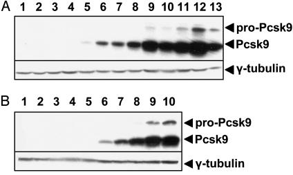
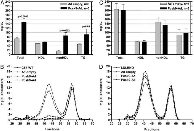
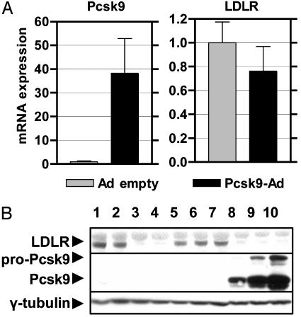
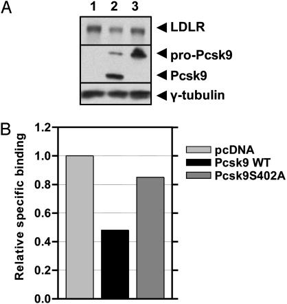
Proprotein convertase subtilisin kexin 9 (Pcsk9) is a subtilisin serine protease with a putative role in cholesterol metabolism. Pcsk9 expression is down-regulated by dietary cholesterol, and mutations in Pcsk9 have been associated with a form of autosomal dominant hypercholesterolemia. To study the function of Pcsk9 in mice, an adenovirus constitutively expressing murine Pcsk9 (Pcsk9-Ad) was used. Pcsk9 overexpression in wild-type mice caused a 2-fold increase in plasma total cholesterol and a 5-fold increase in non-high-density lipoprotein (HDL) cholesterol, with no increase in HDL cholesterol, as compared with mice infected with a control adenovirus. Fast protein liquid chromatography analysis showed that the increase in non-HDL cholesterol was due to an increase in low-density lipoprotein (LDL) cholesterol. This effect appeared to depend on the LDL receptor (LDLR) because LDLR knockout mice infected with Pcsk9-Ad had no change in plasma cholesterol levels as compared with knockout mice infected with a control adenovirus. Furthermore, whereas overexpression of Pcsk9 had no effect on LDLR mRNA levels, there was a near absence of LDLR protein in animals overexpressing Pcsk9. These results were confirmed in vitro by the demonstration that transfection of Pcsk9 in McA-RH7777 cells caused a reduction in LDLR protein and LDL binding. In summary, these results indicate that overexpression of Pcsk9 interferes with LDLR-mediated LDL cholesterol uptake. Because Pcsk9 and LDLR are coordinately regulated by cholesterol, Pcsk9 may be involved in a novel mechanism to modulate LDLR function by an alternative pathway than classic cholesterol inhibition of sterol regulatory element binding protein-mediated transcription.


Figures

Fig. 1.
Fig. 1.
Adenoviral-mediated expression of Pcsk9. (A) Male C57BL/6 mice of >10 weeks of age that were fed a chow diet were injected through the tail vein with PBS (lanes 1 and 2), Ad empty (lanes 3 and 4), or an adenovirus constitutively expressing the murine C57BL/6 Pcsk9 ORF under the control of a CMV promoter (Pcsk9-Ad, lanes 5–12). Pcsk9-Ad was injected at increasing concentrations from 1 × 10⁸ pfu per mouse to 3 × 10⁹ pfu per mouse (lanes 5–12). As a control, McA-RH7777 cells were infected with Pcsk9-Ad at an moi of 2,000 (lane 13). (B) Male LDLR knockout mice on a C57BL/6 background of >10 weeks of age that were fed a chow diet were injected through the tail vein with Ad empty (lanes 1–5) or increasing concentrations of Pcsk9-Ad (lanes 6–10). In both cases, liver homogenates were immunoblotted with an antibody to Pcsk9 and γ-tubulin.
Fig. 2.
Fig. 2.
Overexpression of Pcsk9 increases LDL cholesterol levels in an LDLR-dependent manner. (A) Plasma was isolated from wild-type male C57BL/6 mice injected with Ad empty and Pcsk9-Ad, and HDL and non-HDL plasma fractions were separated by ultracentrifugation. Cholesterol levels in these fractions were measured by enzymatic assay. Overexpression of Pcsk9 caused an increase in total and non-HDL cholesterol with no change in HDL cholesterol levels. (B) Plasma was pooled from at least two wild-type animals (C57 WT), animals injected with Ad empty, and animals injected with Pcsk9-Ad; plasma was fractionated by FPLC; and cholesterol was measured by enzymatic assay. Two runs of Pcsk9-Ad-injected mice are shown to demonstrate reproducibility. Wild-type mice and mice injected with Ad empty had mainly HDL-derived cholesterol (fractions 52–62). Mice injected with Pcsk9-Ad had similar levels of HDL-derived and very-low-density lipoprotein-derived cholesterol, but they had high levels of LDL-derived cholesterol (fractions 35–50). (C) LDLR knockout mice were injected with Ad empty and Pcsk9-Ad. LDLR knockout mice injected with Ad empty had elevated total cholesterol and non-HDL cholesterol with no change in HDL cholesterol levels. Overexpression of Pcsk9 did not cause a further increase in total or non-HDL cholesterol levels in LDLR knockout mice. (D) FPLC analysis demonstrated that the lipoprotein profile of LDLR knockout mice injected with Pcsk9-Ad was indistinguishable from that of uninjected LDLR knockout mice (LDLRKO) and LDLR knockout mice injected with Ad empty.
Fig. 3.
Fig. 3.
Overexpression of Pcsk9 results in an absence of LDLR protein with normal LDLR mRNA levels. (A) The levels of LDLR mRNA in livers of mice injected with Ad empty and Pcsk9-Ad was measured by quantitative RT-PCR. Overexpression of Pcsk9 did not change the levels of LDLR mRNA. (B) The levels of LDLR protein in livers of mice injected with Ad empty and Pcsk9-Ad were detected by immunoblotting with an antibody specific to the murine LDLR cytoplasmic tail. Wild-type C57BL/6 mice (lanes 1 and 2) had a predominant LDLR band at ≈160 kDa; this band was absent in LDLR knockout mice (lanes 3 and 4). Mice injected with Ad empty had normal levels of LDLR protein (lanes 5–7), whereas mice injected with Pcsk9-Ad had a complete absence of LDLR protein (lanes 8–10).
Fig. 4.
Fig. 4.
Overexpression of Pcsk9 in vitro results in a decrease in LDLR protein and LDL binding. (A) McA-RH7777 cells were transiently transfected with pcDNA empty vector (lane 1), wild-type Pcsk9-pcDNA (lane 2), and the mutant Pcsk9(S402A)-pcDNA (lane 3). Overexpression of wild-type Pcsk9 resulted in a 72% decrease in LDLR protein, whereas overexpression of catalytically inactive Pcsk9 caused a 15% decrease in LDLR protein. (B) Specific binding of DiI-LDL was measured in transiently transfected McA-RH7777 cells. Overexpression of wild-type Pcsk9 resulted in a 52% decrease in DiI-LDL binding, whereas overexpression of catalytically inactive Pcsk9 resulted in a 15% decrease in DiI-LDL binding. Data shown are representative of three independent experiments.

References

    1. Hopkins, P. N. (1992) Am. J. Clin. Nutr. 55, 1060-1070. - PubMed
    1. Herron, K. L., Vega-Lopez, S., Conde, K., Ramjiganesh, T., Shachter, N. S. & Fernandez, M. L. (2003) J. Nutr. 133, 1036-1042. - PubMed
    1. Clarke, R., Frost, C., Collins, R., Appleby, P. & Peto, R. (1997) Br. Med. J. 314, 112-117. - PMC - PubMed
    1. Maxwell, K. N., Soccio, R. E., Duncan, E. M., Sehayek, E. & Breslow, J. L. (2003) J. Lipid Res. 44, 2109-2119. - PubMed
    1. Siezen, R. J. & Leunissen, J. A. (1997) Protein Sci. 6, 501-523. - PMC - PubMed

Publication types

LinkOut - more resources