

Review
doi: 10.1016/j.tips.2004.03.010.
K+ channels as targets for specific immunomodulation
Affiliations
- PMID: 15120495
- PMCID: PMC2749963
- DOI: 10.1016/j.tips.2004.03.010
Item in Clipboard
Review
K+ channels as targets for specific immunomodulation
Trends Pharmacol Sci.
2004 May.
Abstract
The voltage-gated Kv1.3 channel and the Ca(2+)-activated IKCa1 K(+) channel are expressed in T cells in a distinct pattern that depends on the state of lymphocyte activation and differentiation. The channel phenotype changes during the progression from the resting to the activated cell state and from naïve to effector memory cells, affording promise for specific immunomodulatory actions of K(+) channel blockers. In this article, we review the functional roles of these channels in both naïve cells and memory cells, describe the development of selective inhibitors of Kv1.3 and IKCa1 channels, and provide a rationale for the potential therapeutic use of these inhibitors in immunological disorders.
Figures
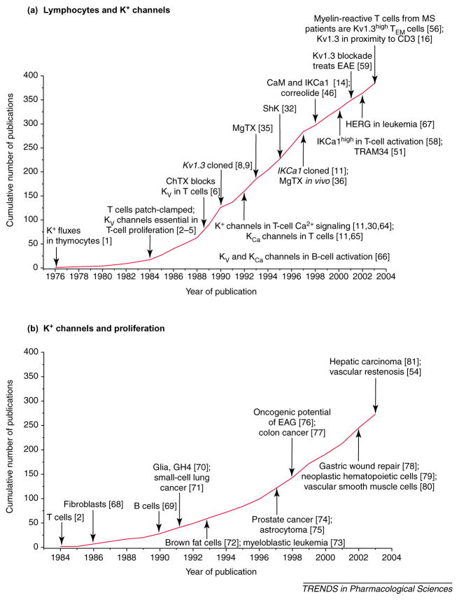
The cumulative number of publications as of November 2003 on (a) ‘lymphocytes and K+ channels’ and (b) ‘K+ channels and proliferation’. Key discoveries in both fields are highlighted. Abbreviations: CaM, calmodulin; ChTX, charybdotoxin; EAE, experimental autoimmune encephalomyelitis; EAG, ether a-go-go; IKCa1, intermediate-conductance Ca2+ -activated K+ channel subtype 1; HERG, human ether-à-go-go-related gene; Kv1.3high, high levels of Kv1.3 channels; MgTX, margatoxin; MS, multiple sclerosis; ShK, Stichodactyla helianthus toxin; TEM cells, effector memory T cells. (Data are from [–,,,,,,,,,,,,,,,,–81].)
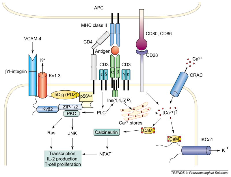
The involvement of voltage-dependent Kv1.3 channels, intermediate-conductance Ca2+ -activated IKCa1 channels and voltage-independent Ca2+ release-activated Ca2+ (CRAC) channels in the activation of a CD4+ T cells by an antigen-presenting cell (APC) is shown. Engagement of the T-cell receptor–CD3 complex through an antigenic peptide presented in the context of major histocompatibility complex (MHC) class II activates phospholipase C (PLC), which leads to the activation of protein kinase C (PKC) and the production of inositol (1,4,5)-trisphosphate [Ins(1,4,5) P3], which liberates Ca2+ from intracellular stores. Simultaneous activation of CD28 by the co-stimulatory molecules CD80 or CD86 further amplifies the resulting Ca2+ signal. The rise in the intracellular concentration of Ca2+ activates the phosphatase calcineurin, which then dephosphorylates the transcription factor nuclear factor of activated T cells (NFAT), enabling it to accumulate in the nucleus and bind to the promoter of the gene encoding interleukin 2 (IL-2). Parallel activation of the c-JUN N-terminal kinase (JNK) and Ras by PKC results in the activation of other transcription factors and initiates transcription of various genes and finally T-cell proliferation. CRAC, Kv1.3 and IKCa1 channels regulate Ca2+ signaling. Depletion of internal Ca2+ stores causes CRAC channels in the membrane to open, and the ensuing Ca2+ influx sustains elevated levels of cytosolic Ca2+. Ca2+ influx through CRAC channels is reduced following membrane depolarization. The driving force for Ca2+ entry is restored by membrane hyperpolarization brought about by the opening of Kv1.3 channels in response to membrane depolarization and the opening of IKCa1 channels as a consequence of elevated concentrations of cytosolic Ca2+. Selective blockade of K+ channels leads to membrane depolarization, inhibits Ca2+ influx and shuts down cytokine production and cell proliferation. Abbreviations: CaM, calmodulin; hDlg, human homolog of the Drosophila discs large tumor suppressor protein; VCAM-4, vascular cell adhesion molecule 4; ZIP-1, Zrt/Irt-like protein.
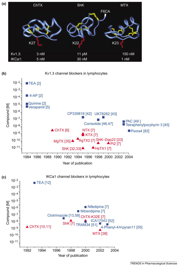
(a) Backbone structures of charybdotoxin (ChTX), Stichodactyla helianthus toxin (ShK) and maurotoxin (MTX), which are peptide inhibitors of voltage-dependent Kv1.3 channels and intermediate-conductance Ca2+ -activated IKCa1 channels. The crucial lysine that occludes the channel pore is highlighted in red in each structure: Lys27 in ChTX; Lys22 in ShK; and Lys23 in MTX. The disulfide bonds are highlighted in yellow. Arg1 in ShK, to which the fluorophore fluorescein-6-carboxylic acid (F6CA) is attached, is highlighted in white. Such fluorophores attached to specific inhibitors can be used to visualize these K+ channels. (b,c) Plots of potency versus year of publication for peptide (red; triangles) and small-molecule (blue; squares) blockers of Kv1.3 (b) and IKCa1 (c) channels in lymphocytes are shown. Abbreviations: AgTX2, agio-toxin-2; 4-AP, 4-aminopyridine; ChTX-K32E, charybdotoxin derivative with glutamate at position 32 in place of the native lysine; HsTX1, Heterometrus spinnifer toxin 1; KTX, kaliotoxin; MgTX, margatoxin; NTX, noxiustoxin; PAC, 4-phenyl-4-[3-(2-methoxyphenyl)-3-oxo-2-azaprop-1-yl]cyclohexanone; Pi2, Pandinus imperator toxin 2; ShK–Dap22, Stichodactyla helianthus toxin with diaminopropionic acid introduced at position 22 in place of the native lysine; TEA, tetraethylammonium chloride. See Chemical names. (Data are from [,–,–,,,,,,,–47,49,51,52,55,58,82].)
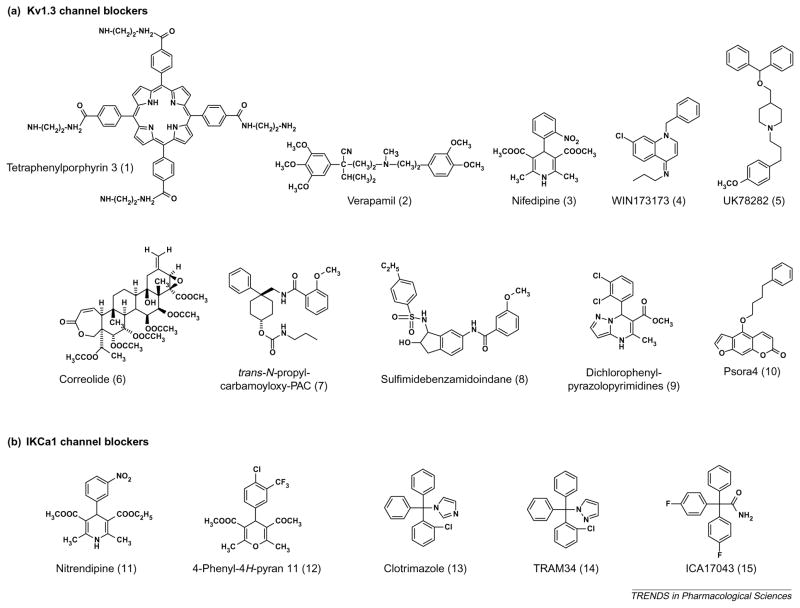
Structures of small-molecule Kv1.3 channel and IKCa1 channel blockers. See Tables 1 and 2 for their Kd values on the respective channels.
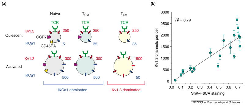
(a) Schematic showing the average numbers of Kv1.3 channels and IKCa1 channels per cell in naïve (CCR7+ CD45RA+ ), central memory (TCM) (CCR7+ CD45RA−) and effector memory (TEM) (CCR7−CD45RA−) cells. Naïve and TCM cells increase IKCa1 channel expression following activation, whereas TEM cells increase Kv1.3 channel expression. (b) Plot of Kv1.3 channel numbers per cell determined by whole-cell patch-clamp in different T-cell populations versus difference (D) values of Stichodactyla helianthus toxin (ShK)–fluorescein-6-carboxylic acid (F6CA) staining obtained by flow cytometry; these data were obtained from [40]. The D value is a measure of the difference in fluorescence intensity between ShK–F6CA-stained cells and background fluorescence from unstained cells of the same population. Abbreviation: TCR, T-cell receptor.
References
-
- Iversen JG. Unidirectional K+ fluxes in rat thymocytes stimulated by concanavalin A. J Cell Physiol. 1976;89:267–276. - PubMed
-
- DeCoursey TE, et al. Voltage-gated K+ channels in human T lymphocytes: a role in mitogenesis? Nature. 1984;307:465–468. - PubMed
-
- Matteson DR, Deutsch C. K+ channels in T lymphocytes: a patch clamp study using monoclonal antibody adhesion. Nature. 1984;307:468–471. - PubMed
Publication types
MeSH terms
Substances
Grants and funding
LinkOut - more resources
Full Text Sources
Other Literature Sources
Molecular Biology Databases
Miscellaneous
