. 2004 May;24(10):4151-65.
doi: 10.1128/MCB.24.10.4151-4165.2004.

The functions of budding yeast Sae2 in the DNA damage response require Mec1- and Tel1-dependent phosphorylation

Affiliations

The functions of budding yeast Sae2 in the DNA damage response require Mec1- and Tel1-dependent phosphorylation

Enrico Baroni et al. Mol Cell Biol. 2004 May.

Abstract

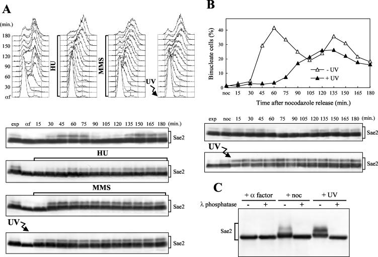
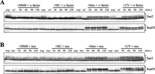
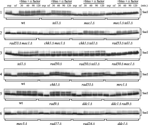
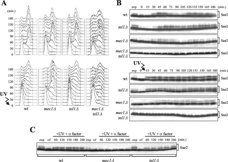
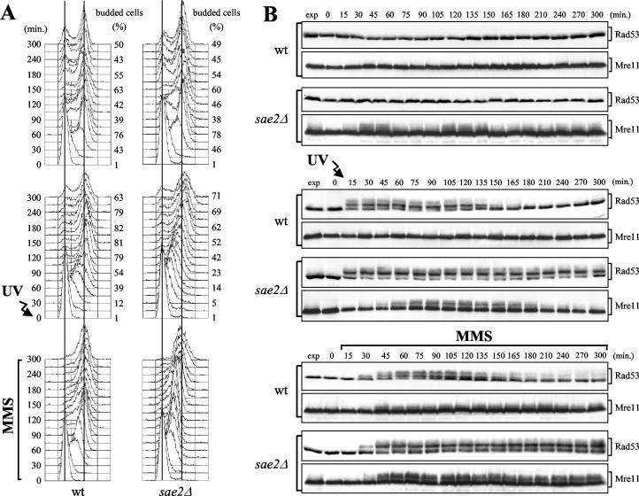
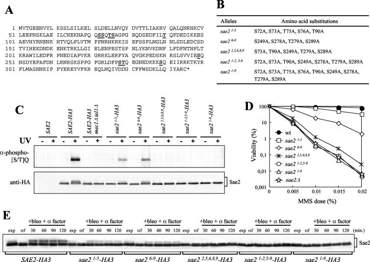
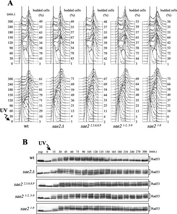
DNA damage checkpoint pathways sense DNA lesions and transduce the signals into appropriate biological responses, including cell cycle arrest, induction of transcriptional programs, and modification or activation of repair factors. Here we show that the Saccharomyces cerevisiae Sae2 protein, known to be involved in processing meiotic and mitotic double-strand breaks, is required for proper recovery from checkpoint-mediated cell cycle arrest after DNA damage and is phosphorylated periodically during the unperturbed cell cycle and in response to DNA damage. Both cell cycle- and DNA damage-dependent Sae2 phosphorylation requires the main checkpoint kinase, Mec1, and the upstream components of its pathway, Ddc1, Rad17, Rad24, and Mec3. Another pathway, involving Tel1 and the MRX complex, is also required for full DNA damage-induced Sae2 phosphorylation, that is instead independent of the downstream checkpoint transducers Rad53 and Chk1, as well as of their mediators Rad9 and Mrc1. Mutations altering all the favored ATM/ATR phosphorylation sites of Sae2 not only abolish its in vivo phosphorylation after DNA damage but also cause hypersensitivity to methyl methanesulfonate treatment, synthetic lethality with RAD27 deletion, and decreased rates of mitotic recombination between inverted Alu repeats, suggesting that checkpoint-mediated phosphorylation of Sae2 is important to support its repair and recombination functions.


Figures

FIG. 1.
FIG. 1.
Sae2 phosphorylation during the unperturbed cell cycle and in response to genotoxic treatments. (A) Exponentially growing YLL1103 cells, expressing Sae2-HA3 from the SAE2 promoter, were synchronized with α-factor (αf) and released from the pheromone block in YEPD, YEPD containing 50 mM HU, or 0.02% MMS, or were UV irradiated (40 J/m2) prior to the release in YEPD. Cell samples collected at the indicated times after α-factor release were analyzed by fluorescence-activated cell sorting (top), and protein extracts were prepared and analyzed by Western blotting with anti-HA antibodies (bottom). (B) Exponentially growing YLL1103 cells were synchronized with nocodazole (noc) and released from the G2 block in YEPD, or were UV irradiated (45 J/m2) prior to the release in YEPD. Cell samples were collected at the indicated times after release from nocodazole to analyze the percentage of binucleate cells by propidium iodide staining (top) and the Sae2-HA3 protein as in panel A (bottom). (C) Protein extracts from α-factor-arrested (+ α factor), nocodazole-arrested (+ noc), or UV-treated exponentially growing (+ UV) YLL1103 cells were immunoprecipitated with anti-HA antibodies. Immunoprecipitates were then incubated at 30°C with (+) or without (−) λ phosphatase before electrophoresis and Western blot analysis using anti-HA antibodies. exp, exponentially growing cells.
FIG. 2.
FIG. 2.
DNA damage-induced Sae2 phosphorylation in G1- and G2-arrested cells. (A) Exponentially growing cell cultures of strain DMP3909/7D, expressing Sae2-HA3 from the SAE2 promoter in a bar1Δ background, were synchronized with α factor (αf) and resuspended in YEPD containing 1 μg of α factor/ml in the presence of either 0.02% MMS (+MMS + α factor), 150 mM HU (+HU + α factor), or 20 mU of bleomycin/ml (+bleo + α factor), or after UV-irradiation (40 J/m2) (+UV + α factor). (B) Exponentially growing cell cultures of strain DMP3909/7D were synchronized with nocodazole (noc) and resuspended in YEPD containing nocodazole in the presence of either 0.02% MMS (+MMS + noc), 150 mM HU (+HU + noc), or 20 mU of bleomycin/ml (+bleo + noc), or after UV irradiation (40 J/m2) (+UV + noc). Protein extracts prepared from cell samples collected at the indicated times were subjected to Western blot analysis with anti-HA (Sae2) and anti-Rad53 (Rad53) antibodies. exp, exponentially growing cells.
FIG. 3.
FIG. 3.
DNA damage-induced Sae2 phosphorylation in checkpoint mutants. Strains expressing Sae2-HA3 from the SAE2 promoter in a bar1Δ background were as follows: wild type (wt) (DMP3909/7D), tel1Δ (DMP3920/16D), mec1Δ sml1Δ (DMP3919/3B), mec1Δ tel1Δ sml1Δ (DMP3919/11A), rad53Δ mec1Δ sml1Δ (DMP4295/10A), chk1Δ mec1Δ sml1Δ (DMP4297/5A), chk1Δ tel1Δ (DMP4296/1D), tel1Δ rad53Δ sml1Δ (DMP4298/2B), rad50Δ (DMP4048/7D), rad50Δ tel1Δ (DMP4050/17A), rad50Δ mec1Δ sml1Δ (DMP4052/18A), chk1Δ (DMP4141/12C), rad53Δ sml1Δ (DMP4106/27A), mrc1Δ (DMP4105/3B), rad9Δ (DMP4049/5C), ddc1Δ (YLL1345.6), ddc1Δ rad9Δ (YLL1346.2), mec3Δ (DMP4138/7C), rad17Δ (DMP4137/1D), and rad24Δ (DMP4137/17A). Cell cultures were arrested in G1 with α-factor and then transferred in YEPD containing 20 mU of bleomycin/ml and 1 μg of α-factor/ml (+bleo + α factor). Time zero (αf) corresponds to cell samples taken immediately before bleomycin addition. Protein extracts prepared from cell samples collected at the indicated times were subjected to Western blot analysis with anti-HA antibodies. exp, exponentially growing cells.
FIG. 4.
FIG. 4.
Cell cycle- and DNA damage-dependent Sae2 phosphorylation in the absence of Mec1 and/or Tel1. Strains expressing SAE2-HA3 from the SAE2 promoter were as follows: wild type (YLL1103), tel1Δ (DMP3807/2D), mec1Δ sml1Δ (DMP3806/2A), and mec1Δ tel1Δ sml1Δ (DMP3916/5B). (A and B) Cell cultures growing logarithmically in YEPD were arrested in G1 with α-factor and released from the pheromone block at time zero in YEPD, or were UV-irradiated (40 J/m2) prior to the release in YEPD. Samples of untreated and UV-treated cell cultures were withdrawn at the indicated times after α-factor release in order to analyze the DNA content by fluorescence-activated cell sorting in nonirradiated (A, top) and UV-irradiated (A, bottom) cell cultures and to analyze Sae2 phosphorylation by Western blot analysis with anti-HA antibodies of extracts from nonirradiated (B, top) and UV-irradiated (B, bottom) cell cultures. (C) Cell cultures were synchronized with α-factor and transferred in YEPD containing α factor after UV irradiation (40 J/m2) (+UV + α factor). Time zero (αf) corresponds to cell samples taken immediately before UV treatment. Protein extracts from samples withdrawn at the indicated times were treated as described for panel B. exp, exponentially growing cells.
FIG. 5.
FIG. 5.
Checkpoint response to DNA damage in sae2Δ cells. Exponentially growing wild-type (wt) (YLL1072.1) and sae2Δ (DMP4224/5B) cell cultures were synchronized with α factor and released from the pheromone block either in YEPD without any treatment (top), in YEPD after UV-irradiation (30 J/m2) (center), or in YEPD containing 0.01% MMS (bottom). Samples were withdrawn at the indicated times after α-factor release (time zero) in order to analyze the DNA content by fluorescence-activated cell sorting and the percentage of budded cells (A) and to monitor Rad53 and Mre11 phosphorylation by Western blot analysis with anti-Rad53 and anti-HA antibodies, respectively (B). exp, exponentially growing cells.
FIG. 6.
FIG. 6.
Substitutions of S or T to A at putative Sae2 phosphorylation sites affect Sae2 phosphorylation and cell survival after MMS treatment. (A) Sae2 amino acid sequence. Numbers on the left indicate the position of the first amino acid residue in each row. The S or T putative phospho-acceptor residues are boldfaced and underlined. (B) Mutant sae2 alleles obtained by site-directed mutagenesis are listed, together with the resulting serine- or threonine-to-alanine changes. (C) Strains were as follows: SAE2 (YLL1348.1), SAE2-HA3 (YLL1351.8), SAE2-HA3 mec1Δ tel1Δ sml1Δ (DMP3916/5B), sae21-5-HA3 (YLL1359.4), sae26-9-HA3 (YLL1360.31), sae22,5,6,8,9-HA3 (YLL1405), sae21-2,5-9-HA3 (YLL1361.44), and sae21-9-HA3 (YLL1352.2). Immunoprecipitations with anti-HA antibodies were performed on protein extracts prepared from nonirradiated (−) or UV-irradiated (+) α-factor-arrested cells of the indicated genotypes. Immunoprecipitates were subjected to Western blot analysis using polyclonal rabbit anti-phosphorylated-(S/T)Q antibodies (α-phospho-[S/T]Q) and anti-HA antibodies. (D) Dose-response killing curves were determined by plating serial dilutions of wild-type (YLL1348.1), sae2Δ (YLL1069.3), sae21-5 (YLL1353.1), sae26-9 (YLL1354.1), sae22,5,6,8,9 (YLL1395.1), sae21-2,5-9 (YLL1355.1), and sae21-9 (YLL1347.1) cell cultures, growing exponentially in YEPD, onto YEPD plates with or without MMS at the indicated concentrations. Plates were incubated at 26°C, and CFU were counted after 3 days. (E) α-factor-arrested cultures of the strains listed in the legend to panel C were transferred to YEPD medium containing 20 mU of bleomycin/ml and 1 μg of α factor/ml (+bleo + α factor). Time zero (αf) corresponds to cell samples taken immediately before bleomycin addition. Protein extracts prepared from cell samples collected at the indicated times were subjected to Western blot analysis with anti-HA antibodies. exp, exponentially growing cells.
FIG. 7.
FIG. 7.
DNA damage checkpoint response in sae2 phosphorylation-defective mutants. Exponentially growing wild-type (K699), sae2Δ (YLL1069.3), sae22,5,6,8,9 (YLL1395.1), sae21-2,5-9 (YLL1355.1), and sae21-9 (YLL1347.1) cell cultures, growing exponentially in YEPD, were synchronized with α factor and released from the pheromone block in YEPD (A, top), or were UV irradiated (30 J/m2) (A, bottom) prior to the release in YEPD. Samples were withdrawn at the indicated times after α-factor release (time zero) in order to analyze the DNA content by fluorescence-activated cell sorting and determine the percentage of budded cells (A) and to detect Rad53 by Western blot analysis with anti-Rad53 antibodies in UV-irradiated cell extracts (B). exp, exponentially growing cells.

Similar articles

Cited by

References

    1. Abraham, R. T. 2001. Cell cycle checkpoint signaling through the ATM and ATR kinases. Genes Dev. 15:2177-2196. - PubMed
    1. Alcasabas, A. A., A. J. Osborn, J. Bachant, F. Hu, P. J. Werler, K. Bousset, K. Furuya, J. F. Diffley, A. M. Carr, and S. J. Elledge. 2001. Mrc1 transduces signals of DNA replication stress to activate Rad53. Nat. Cell Biol. 3:958-965. - PubMed
    1. Aylon, Y., and M. Kupiec. 2003. The checkpoint protein Rad24 of Saccharomyces cerevisiae is involved in processing double-strand break ends and in recombination partner choice. Mol. Cell. Biol. 23:6585-6596. - PMC - PubMed
    1. Bashkirov, V. I., J. S. King, E. V. Bashkirova, J. Schmuckli-Maurer, and W. D. Heyer. 2000. DNA repair protein Rad55 is a terminal substrate of the DNA damage checkpoints. Mol. Cell. Biol. 20:4393-4404. - PMC - PubMed
    1. Bentley, N. J., D. A. Holtzman, G. Flaggs, K. S. Keegan, A. DeMaggio, J. C. Ford, M. Hoekstra, and A. M. Carr. 1996. The Schizosaccharomyces pombe rad3 checkpoint gene. EMBO J. 15:6641-6651. - PMC - PubMed

Publication types

MeSH terms