. 2004 May;24(10):4351-60.
doi: 10.1128/MCB.24.10.4351-4360.2004.

Protein kinase C-alpha phosphorylation of specific serines in the connecting segment of the beta 4 integrin regulates the dynamics of type II hemidesmosomes

Affiliations

Protein kinase C-alpha phosphorylation of specific serines in the connecting segment of the beta 4 integrin regulates the dynamics of type II hemidesmosomes

Isaac Rabinovitz et al. Mol Cell Biol. 2004 May.

Abstract

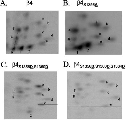
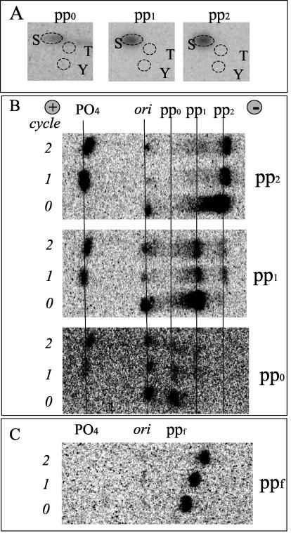
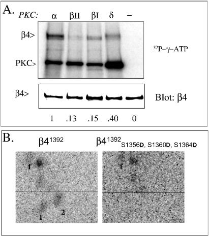
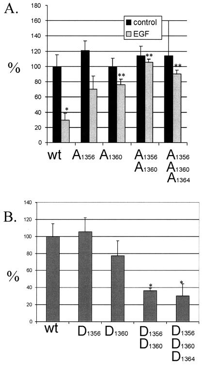
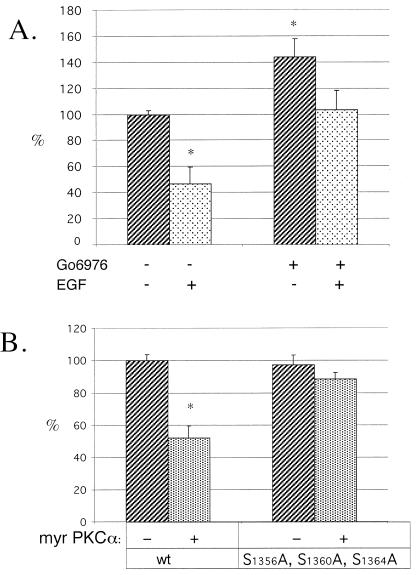
Although the regulation of hemidesmosome dynamics during processes such as epithelial migration, wound healing, and carcinoma invasion is important, the mechanisms involved are poorly understood. The integrin alpha 6 beta 4 is an essential component of the hemidesmosome and a target of such regulation. Epidermal growth factor (EGF) can induce hemidesmosome disassembly by a mechanism that involves serine phosphorylation of the beta 4 integrin subunit. Using a combination of biochemical and mutational analyses, we demonstrate that EGF induces the phosphorylation of three specific serine residues (S(1356), S(1360), and S(1364)) located within the connecting segment of the beta 4 subunit and that phosphorylation on these residues accounts for the bulk of beta 4 phosphorylation stimulated by EGF. Importantly, phosphorylation of these serines is critical for the ability of EGF to disrupt hemidesmosomes. Using COS-7 cells, which assemble hemidesmosomes type II upon exogenous expression of the alpha 6 beta 4 integrin, we observed that expression of a beta 4 construct containing Ser-->Ala mutations of S(1356), S(1360), and S(1364) reduced the ability of EGF to disrupt hemidesmosomes and that this effect appears to involve cooperation among these phosphorylation sites. Moreover, expression of Ser-->Asp mutants that mimic constitutive phosphorylation reduced hemidesmosome formation. Protein kinase C-alpha (PKC-alpha) is the kinase responsible for phosphorylating at least two of these serines, based on in vitro kinase assays, peptide mapping, and mutational analysis. Together, these results highlight the importance of serine phosphorylation in regulating type II hemidesmosome disassembly, implicate a cluster of serine residues within the connecting segment of beta 4, and argue for a key role for PKC-alpha in regulating these structures.


Figures

FIG. 1.
FIG. 1.
EGF stimulates phosphorylation of the β4 integrin largely on serine. (A) HaCat cells plated on laminin-1 were labeled with 32PO4 as described in Materials and Methods and stimulated with EGF (100 ng/ml) for 15 min. Cells were lysed, and the α6β4 integrin was immunoprecipitated, separated using SDS-PAGE, blotted with the β4-specific polyclonal antibody, and exposed to a phosphor screen. (B) Phosphoamino acid analysis of 32PO4-labeled β4. The radiolabeled band that corresponded to the β4 subunit obtained from cells stimulated with either 0 (−) or 100-ng/ml (+) EGF was cut from the membrane, subjected to acid hydrolysis, and resolved using two-dimensional TLE as described in Materials and Methods. The plate was then exposed to a phosphor screen. S, phosphoserine; T, phosphothreonine; Y, phosphotyrosine.
FIG. 2.
FIG. 2.
Peptide mapping analysis of phosphorylated wild-type and mutant β4 integrin. Excised bands containing 32P-radiolabeled wild-type β4 from HaCat cells (A) or mutated β4 from COS-7 transfectants (B to D) were digested using trypsin, and the peptides were resolved in two dimensions using a combination of TLE and TLC and exposed to a phosphor screen. The phosphopeptides of interest for this study (pp0, pp1, and pp2) are located below a line defined by a control dye. Notice the disappearance of pp0 and both an increase and relocation of pp1 and pp2 to more hydrophobic (higher) positions in β4S1356A (B); the disappearance of pp0 and pp1, as well as the relocation of pp2 to a more hydrophilic (lower) position in the double mutant β4S1356D S1360D (C); and the disappearance of pp0, pp1, and pp2 in the triple mutant β4S1356D S1360D S1364D (D).
FIG. 3.
FIG. 3.
Analysis of phosphorylated peptides (pp0, pp1, and pp2) derived from radiolabeled β4 integrin. 32P radiolabeled peptides were eluted from TLE or TLC plates and subjected to phosphoamino acid analysis (A) or Edman degradation (B and C). The peptides were degraded for at least 2 cycles. Notice that the phosphate is shed during the first cycle for pp0, pp1, and pp2 (B). In contrast, ppf (C), used as a control, did not shed any phosphate (PO4 position) during the first 2 cycles. ori, sample origin; +, anode; −, cathode; S, phosphoserine; T, phosphothreonine; Y, phosphotyrosine.
FIG. 4.
FIG. 4.
Identification of S1356, S1360, and S1364 as the major sites of β4 phosphorylation. (A) Two candidate peptides (PP-I and PP-II) were identified based on a combined analysis that included phosphopeptide mobility, Edman degradation, phosphoamino acid analysis, and diagnostic enzymatic digestion. (B) Point mutations introduced into the candidate serines in the full-length β4 were analyzed by peptide mapping as described in Fig. 2. The table notes the presence or disappearance of the three major peptides of interest after peptide mapping. The peptide maps of mutants critical for the identification process are shown on Fig. 2. (C) Position of S1356, S1360, and S1364 within the β4 integrin cytoplasmic tail. FNn, fibronectin type III repeats; CS, connecting segment. (D) Structure of β4 integrin point mutations (S1356, S1360, and S1364) constructed on a full-length β4 for this study. Two sets of mutants were built: Ser→Ala, to block phosphorylation and Ser→Asp to mimic constitutive phosphorylation.
FIG. 5.
FIG. 5.
PKC-α phosphorylates pp1 and pp2 in vitro. Lysates were obtained from COS-7 cells transfected with wild-type β4 (A), truncated β41392 (B, left) or triple Ser→Asp mutant β41392 S1356D S1360D S1364D (B, right) were immunocomplexed with anti-β4 antibodies and antirat agarose and labeled with [γ-32P]ATP and either PKC-α, -βΙ, -βΙΙ, or -δ as described in Materials and Methods. Radiolabeled immunocomplexes were eluted and separated by SDS-PAGE, electrotransferred to a PVDF membrane, and exposed to a phosphor screen (A, top panel) and later blotted with an anti-β4 polyclonal antibody (A, bottom panel) Autophosphorylation of PKC indicates the activity of the enzymes used in the assay. In panel B, radiolabeled β4 was excised from the PVDF membrane, trypsinized, and analyzed by peptide mapping as described in the legend to Fig. 2.
FIG. 6.
FIG. 6.
Expression of hemidesmosomal plaques in COS-7 transfectants. Examples of COS-7 transfectants determined to be positive (A, B, C) or negative (D, E, and F) for expression of hemidesmosomal plaques. Panels A to C show COS-7 cells transfected with wild-type β4 + α6 integrin subunits. Panels C to E show COS-7 cells transfected with β4S1356D S1360D S1364D + α6 integrin subunits. The cells were grown overnight on coverslips and processed for indirect immunofluorescence. The cells were stained for HD1/plectin (B and E [red in panel C or F]) and α6β4 (A and D [green in panel C or F]) and analyzed for colocalization (yellow in panel C or F). Notice the hemidesmosomal plaques in panel C and only a thread-like pattern in panel F. Bar, 10 μm.
FIG. 7.
FIG. 7.
Quantitation of the effect of serine mutations on hemidesmosome dynamics. (A and B) COS-7 cells transfected with the indicated Ser→Ala (A) or Ser→Asp (B) phosphorylation mutants as described in Materials and Methods. (A) The Ser→Ala mutants were incubated in the absence or presence of EGF (100 ng/ml) overnight and processed for indirect immunofluorescence as described in the legend to Fig. 6 and Materials and Methods. The graph describes the percentage of positive cells showing the presence of hemidesmosomal plaques in relation to the wild type β4 (wt; 100%). (B) The Ser→Asp mutants were incubated overnight on coverslips and then processed for indirect immunofluorescence and analyzed as described for panel A. *, P < 0.05 versus wild type; **, P < 0.05 versus wild type + EGF.
FIG. 8.
FIG. 8.
β4 serines S1356, S1360, and S1364 mediate the effects of PKC-α on hemidesmosomal plaques. (A). COS-7 cells expressing exogenous α6β4 were incubated overnight on coverslips in the presence or absence of Gö6976 and/or EGF and then analyzed and processed for indirect immunofluorescence as described in the legend to Fig. 6 and Materials and Methods. The graph describes the percentage of positive cells showing the presence of hemidesmosomal plaques in relation to the unstimulated cells (100%). (B) COS-7 cells were cotransfected with α6, β4, and myrPKC-α. The cells were plated on coverslips overnight and processed and analyzed as described for panel A.

References

    1. Alt, A., M. Ohba, L. Li, M. Gartsbein, A. Belanger, M. F. Denning, T. Kuroki, S. H. Yuspa, and T. Tennenbaum. 2001. Protein kinase Cdelta-mediated phosphorylation of alpha6beta4 is associated with reduced integrin localization to the hemidesmosome and decreased keratinocyte attachment. Cancer Res. 61:4591-4598. - PubMed
    1. Beaulieu, J. F. 1997. Extracellular matrix components and integrins in relationship to human intestinal epithelial cell differentiation. Prog. Histochem. Cytochem. 31:1-78. - PubMed
    1. Borradori, L., P. J. Koch, C. M. Niessen, S. Erkeland, M. R. van Leusden, and A. Sonnenberg. 1997. The localization of bullous pemphigoid antigen 180 (BP180) in hemidesmosomes is mediated by its cytoplasmic domain and seems to be regulated by the beta4 integrin subunit. J. Cell Biol. 136:1333-1347. - PMC - PubMed
    1. Borradori, L., and A. Sonnenberg. 1999. Structure and function of hemidesmosomes: more than simple adhesion complexes. J. Investig. Dermatol. 112:411-418. - PubMed
    1. Boyle, W. J., P. van der Geer, and T. Hunter. 1991. Phosphopeptide mapping and phosphoamino acid analysis by two-dimensional separation on thin-layer cellulose plates. Methods Enzymol. 201:110-149. - PubMed

Publication types

MeSH terms