. 2004 May;24(10):4502-12.
doi: 10.1128/MCB.24.10.4502-4512.2004.

TRAF family proteins link PKR with NF-kappa B activation

Affiliations

TRAF family proteins link PKR with NF-kappa B activation

Jesús Gil et al. Mol Cell Biol. 2004 May.

Abstract

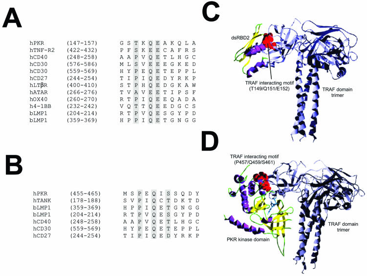
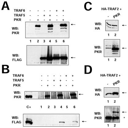
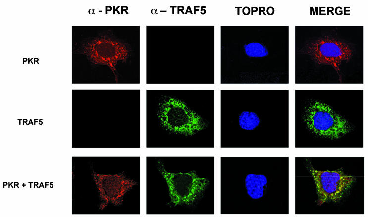
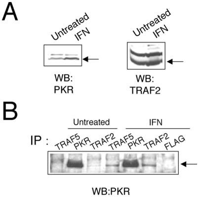
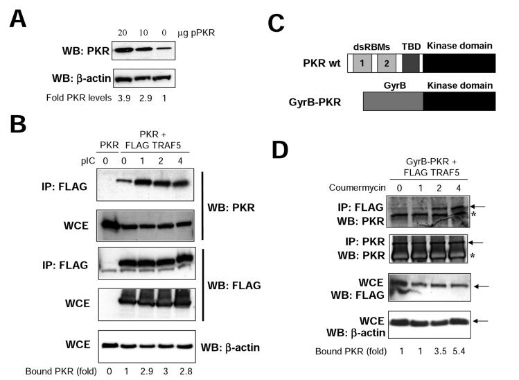
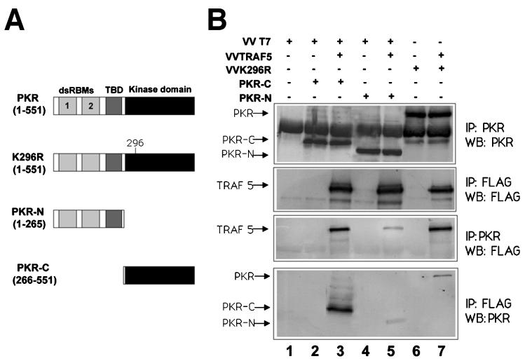
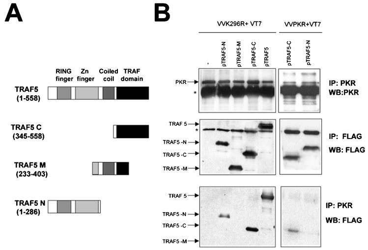
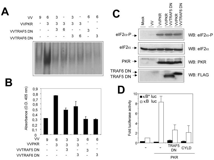
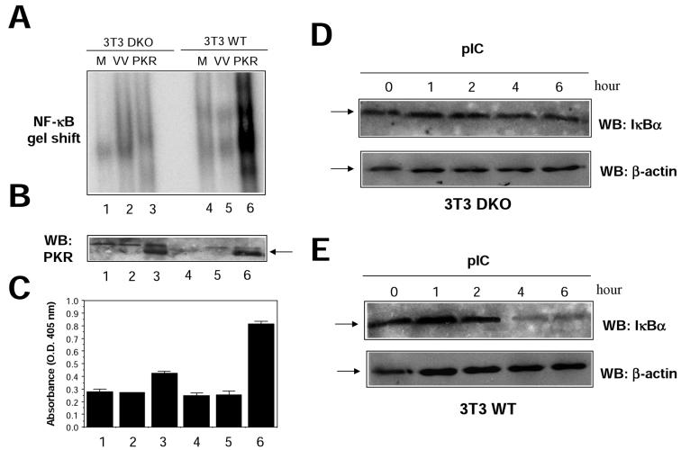
The double-stranded RNA (dsRNA)-dependent protein kinase PKR activates NF-kappa B via the I kappa B kinase (IKK) complex, but little is known about additional molecules that may be involved in this pathway. Analysis of the PKR sequence enabled us to identify two putative TRAF-interacting motifs. The viability of such an interaction was further suggested by computer modeling. Here, we present evidence of the colocalization and physical interaction between PKR and TRAF family proteins in vivo, as shown by immunoprecipitation and confocal microscopy experiments. This interaction is induced upon PKR dimerization. Most importantly, we show that the binding between PKR and TRAFs is functionally relevant, as observed by the absence of NF-kappa B activity upon PKR expression in cells genetically deficient in TRAF2 and TRAF5 or after expression of TRAF dominant negative molecules. On the basis of sequence information and mutational and computer docking analyses, we favored a TRAF-PKR interaction model in which the C-terminal domain of TRAF binds to a predicted TRAF interaction motif present in the PKR kinase domain. Altogether, our data suggest that TRAF family proteins are key components located downstream of PKR that have an important role in mediating activation of NF-kappa B by the dsRNA-dependent protein kinase.


Figures

FIG. 1.
FIG. 1.
Model predicting TRAF-PKR interaction. (A) Sequence alignment of the putative TRAF-interacting motif present in the dsRBD2 subdomain of PKR with those from different TRAF binding domains. (B) Sequence alignment of the putative TRAF-interacting motif present in the kinase domain of PKR with those from different TRAF binding domains. (C) Putative model of the interaction between dsRDB2 and TRAF. A ribbon plot of the structure obtained through docking procedures for the interaction between the TRAF-interacting motif (T149/Q151/E152; red spheres) of the dsRDB2 subdomain of PKR and TRAF. TRAF trimer secondary-structure elements are depicted in grey. The dsRBD amino-terminal domain of PKR is shown as a ribbon plot (α helices, magenta; β strands, yellow). (D) Model of the PKR (kinase domain)-TRAF interaction. A docking model of the putative interaction between the TRAF-interacting motif (P457/Q459/S46; red spheres) of the kinase domain of PKR and the TRAF domain. TRAF trimer secondary-structure elements are depicted in grey. In the kinase domain of PKR, α helices are in magenta and β strands are in yellow. A molecule of ATP has been included solely to indicate the position of the putative ATP binding pocket.
FIG. 2.
FIG. 2.
PKR interacts with TRAF family proteins in vivo. (A) HeLa cells were infected with 10 PFU of VT7 per cell. After 1 h, plasmids encoding FLAG-tagged TRAF5 and TRAF6 proteins or the empty vector were transfected together with the empty vector or a plasmid encoding PKR (K296R mutant form). Cell extracts were collected 20 hpi and analyzed by SDS-PAGE, followed by immunoblot (Western blot [WB]) analysis with anti-PKR or anti-FLAG antiserum. (B) Extracts prepared as described for panel A were immunoprecipitated with anti-PKR serum and thoroughly washed, and immunocomplexes were analyzed by SDS-PAGE and subjected to immunoblotting with antiserum to PKR or FLAG. (C) HeLa cells in 10-cm-diameter plates were infected with 10 PFU of VT7 per cell. After 1 h, a plasmid (10 μg) encoding HA-tagged TRAF2 was transfected together with 10 μg of the empty vector (lane 1) or with 10 μg of a plasmid encoding PKR (K296R mutant form, lane 2). Cells extracts were collected at 20 hpi and analyzed by SDS-PAGE, followed by immunoblot analysis with anti-PKR or anti-HA antiserum. (D) Extracts prepared as described for panel C were immunoprecipitated with anti-HA serum and thoroughly washed, and immunocomplexes were analyzed by SDS-PAGE and subjected to immunoblotting with antiserum to PKR or HA. The asterisk denotes IgGs, and the arrows indicate the specific proteins.
FIG. 3.
FIG. 3.
PKR and TRAF5 colocalize in HeLa cells. HeLa cells were infected at 3 PFU per cell with VV PKR K296R (upper panels), VV FLAG-tagged TRAF5 (middle panels), or both (lower panels). Cells were fixed at 16 hpi and processed for immunofluorescence analysis by confocal microscopy with antibodies directed against PKR (red), FLAG (green), and To-Pro for nuclear staining (blue). Merged images are presented on the right.
FIG. 4.
FIG. 4.
Interaction between PKR and endogenous TRAF proteins. (A) Extracts (50 μg) from HeLa cells left untreated or treated with IFN-α/β for 16 h were separated by SDS-PAGE and analyzed by immunoblotting (Western blotting [WB]) with anti-PKR or anti-TRAF2 antibodies. (B) Extracts (1 mg) were immunoprecipitated (IP) with antibodies against TRAF2, TRAF5, PKR, or FLAG and thoroughly washed, and immunocomplexes were analyzed by SDS-PAGE and subjected to immunoblotting with antiserum to PKR.
FIG. 5.
FIG. 5.
TRAF-PKR interaction is inducible. (A) 293T cells growing in 10-cm-diameter dishes were transfected with 10 or 20 μg of a plasmid coding for PKR, and at 48 h after transfection, cell extracts were collected and analyzed by immunoblotting (Western blotting [WB]) with antiserum to PKR or actin. Signals were quantitated with NIH Image software. (B) 293T cells were transfected with 20 μg of a plasmid coding for PKR (lane 0) or with 10 μg each of plasmids encoding for PKR and FLAG-TRAF5, as indicated. At 48 h after transfection, cells were treated with 10 μg of pIC per ml. At the noted times, cell extracts were collected, immunoprecipitated (IP) with anti-FLAG serum, and thoroughly washed and immunocomplexes or whole-cell extracts (WCE) were analyzed by SDS-PAGE and subjected to immunoblotting with antiserum to PKR, FLAG, or actin. Signals were quantitated with NIH Image software. (C) Schematic representation of PKR and the GyrB-PKR chimera. The different domains of PKR are shown (dsRBMs, the third basic domain [TBD], and the kinase domain). The chimera consists of the N-terminal 220 aa of E. coli GyrB fused to the kinase domain (aa 258 to 551) of human PKR. (D) 293T cells were transfected with 10 μg each of plasmids encoding Gyr-PKR and FLAG-TRAF5 as indicated. At 48 h after transfection, cells were treated with coumermycin. At the indicated times posttreatment, cell extracts were collected, immunoprecipitated with anti-FLAG or anti-PKR serum, and thoroughly washed and immunocomplexes or whole-cell extracts (WCE) were analyzed by SDS-PAGE and subjected to immunoblotting with antiserum to PKR, FLAG, or actin. Asterisks denote IgGs, and arrows indicate the specific proteins. Signals were quantitated with NIH Image software.
FIG. 6.
FIG. 6.
TRAF5 preferentially interacts with the PKR kinase domain. (A) Schematic representation of PKR and the different mutant proteins used. Domains are as in Fig. 5C. (B) HeLa cells grown in 10-cm-diameter plates were infected with 5 PFU of VT7 per cell plus 5 PFU of VV per cell (lanes 1, 2, and 4), 5 PFU of VT7 per cell plus 5 PFU of VV TRAF5 per cell (lanes 3 and 5), 5 PFU of VV per cell plus 5 PFU of VV K296R per cell (lane 6), or 5 PFU of VV TRAF5 per cell plus 5 PFU of VV K296R per cell (lane 7). After 1 h, plasmids encoding PKR-C (10 μg) or PKR-N (10 μg) were transfected as indicated. Cell extracts were collected at 20 hpi, immunoprecipitated (IP) with anti-PKR or anti-FLAG serum, and thoroughly washed, and immunocomplexes were analyzed by SDS-PAGE and subjected to immunoblotting (Western blotting [WB]) with antiserum to PKR or FLAG. TBD, third basic domain.
FIG. 7.
FIG. 7.
PKR preferentially interacts with the TRAF domain of TRAF5. (A) Schematic representation of TRAF5 and the different mutant proteins used. (B) HeLa cells grown in 10-cm-diameter plates were infected with 5 PFU of VT7 per cell and 5 PFU of VV K296R or VV PKR per cell as indicated. After 1 h, 10 μg of a plasmid encoding FLAG-tagged TRAF5 (full-length protein or deletion mutant forms) or 10 μg of the empty vector was transfected. Cell extracts were collected at 20 hpi, immunoprecipitated (IP) with anti-PKR or anti-FLAG serum, and thoroughly washed, and immunocomplexes were analyzed by SDS-PAGE and subjected to immunoblotting (Western blotting [WB]) with antiserum to PKR or FLAG. The asterisks indicate IgGs.
FIG. 8.
FIG. 8.
Dominant negative TRAF proteins inhibit PKR-induced NF-κB activity. (A) HeLa cells were infected with VV recombinants at the numbers of PFU per cell indicated. Nuclear extracts were prepared at 20 hpi and analyzed by gel shift assay with a probe specific for NF-κB. This experiment was performed in duplicate. (B) NF-κB activation was measured in HeLa cells infected with the indicated VV recombinants by using an ELISA as described in Materials and Methods. O.D., optical density. (C) Western blot (WB) analysis of cell extracts from HeLa cells mock infected (first lane) or infected with the indicated viruses was performed with antibodies directed against total or phosphorylated eIF-2a, PKR, or FLAG. (D) 293T cells grown in 10-cm-diameter plates were transfected with equal amounts (10 μg each) of the indicated plasmids together with 0.5 μg of the p3κB-Luc vector (white bars) or the p3κB*Luc vector (with mutated κB sites, black bars). At 36 h after transfection, cells were treated with 10 μg of pIC per ml for 6 h. Luciferase activity was measured, and fold luciferase activity is shown (readings from cells transfected with the empty vector were used as a reference).
FIG. 9.
FIG. 9.
Absence of PKR-dependent NF-κB activation in 3T3 cells deficient in TRAF2 and TRAF5. (A) 3T3 cells grown in 10-cm-diameter plates, obtained from WT mice (WT 3T3) or mice deficient in both TRAF2 and TRAF5 (DKO 3T3), were mock infected (M) or infected with 5 PFU of VV per cell (VV) or 5 PFU of VV PKR per cell (PKR). Nuclear extracts were prepared at 20 hpi and analyzed by gel shift assay with a probe specific for NF-κB. (B) Extracts (50 μg) from the same cells were analyzed for PKR expression by Western blotting (WB). (C) Cells infected as described for panel A were analyzed for induction of apoptosis as indicated in Materials and Methods. The lane numbers in panels B and C are the same as those in panel A. O.D., optical density. DKO (D) or WT (E) 3T3 cells were treated with 10 μg of pIC per ml for the times indicated, and levels of IκBα and β-actin were analyzed by Western blotting. The arrows indicate specific proteins.

References

    1. Akiba, H., H. Nakano, S. Nishinaka, M. Shindo, T. Kobata, M. Atsuta, C. Morimoto, C. F. Ware, N. L. Malinin, D. Wallach, H. Yagita, and K. Okumura. 1998. CD27, a member of the tumor necrosis factor receptor superfamily, activates NF-κB and stress-activated protein kinase/c-Jun N-terminal kinase via TRAF2, TRAF5 and NF-κB-inducing kinase. J. Biol. Chem. 273:13353-13358. - PubMed
    1. Alexopoulou, L., A. C. Holt, R. Medzhitov, and R. A. Flavell. 2001. Recognition of double-stranded RNA and activation of NF-κB by Toll-like receptor 3. Nature 413:732-738. - PubMed
    1. Arch, R. H., R. W. Gedrich, and C. B. Thompson. 1998. Tumor necrosis factor receptor-associated factors (TRAFs)—a family of adapter proteins that regulates life and death. Genes Dev. 12:2821-2830. - PubMed
    1. Arenzana-Seisdedos, F., J. Thompson, M. S. Rodriguez, F. Bachelerie, D. Thomas, and R. T. Hay. 1995. Inducible nuclear expression of newly synthesized IκBα negatively regulates DNA-binding and transcriptional activities of NF-κB. Mol. Cell. Biol. 15:2689-2696. - PMC - PubMed
    1. Bonnet, M. C., R. Weil, E. Dam, A. G. Hovanessian, and E. F. Meurs. 2000. PKR stimulates NF-κB irrespective of its kinase function by interacting with the IκB kinase complex. Mol. Cell. Biol. 20:4532-4542. - PMC - PubMed

Publication types

MeSH terms