. 2004 Apr 30;32(8):2396-410.
doi: 10.1093/nar/gkh527. Print 2004.

DNA binding and antigene activity of a daunomycin-conjugated triplex-forming oligonucleotide targeting the P2 promoter of the human c-myc gene

Affiliations

DNA binding and antigene activity of a daunomycin-conjugated triplex-forming oligonucleotide targeting the P2 promoter of the human c-myc gene

Giuseppina M Carbone et al. Nucleic Acids Res. .

Abstract

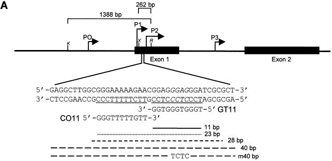
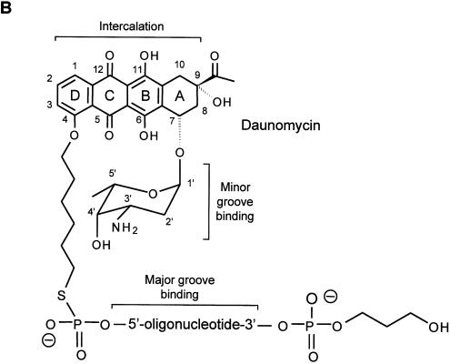
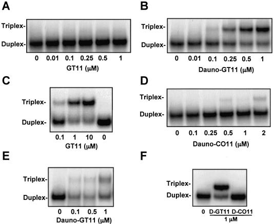
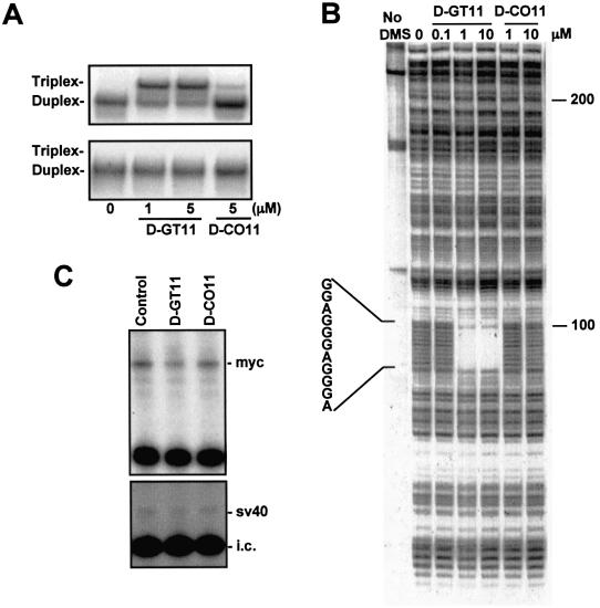
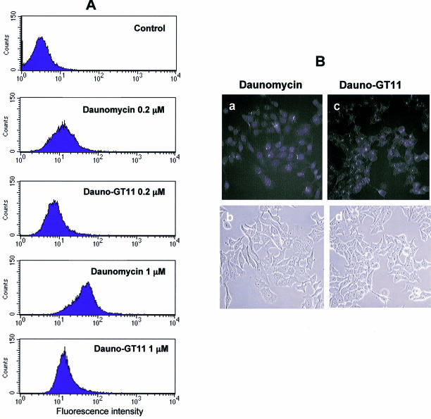
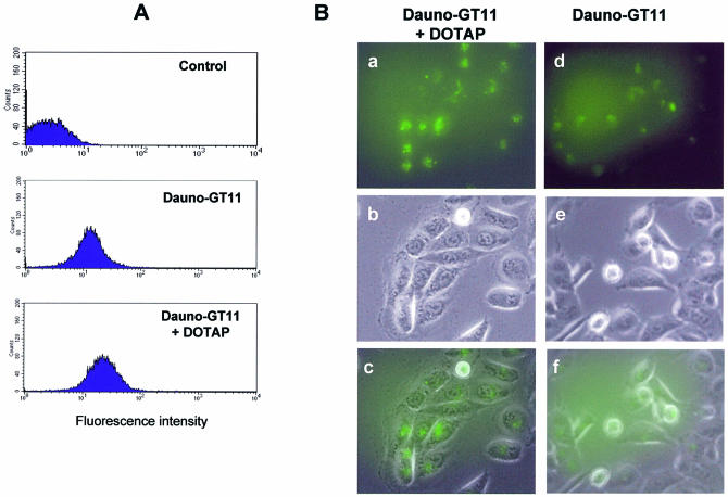
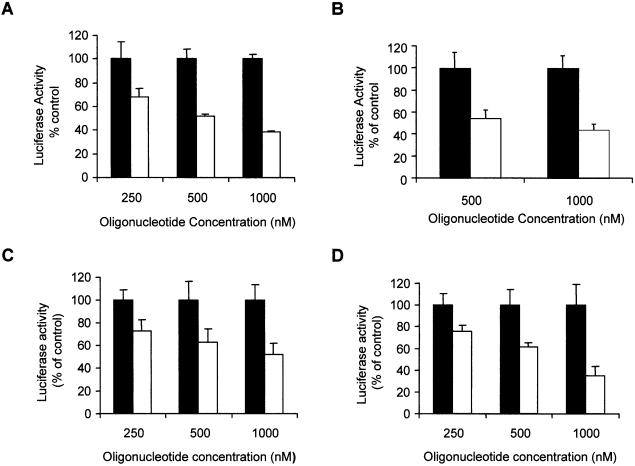
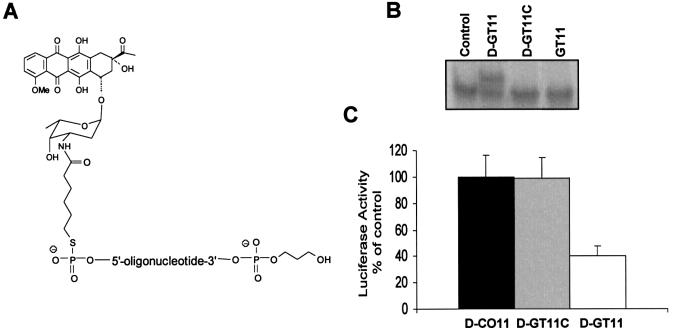
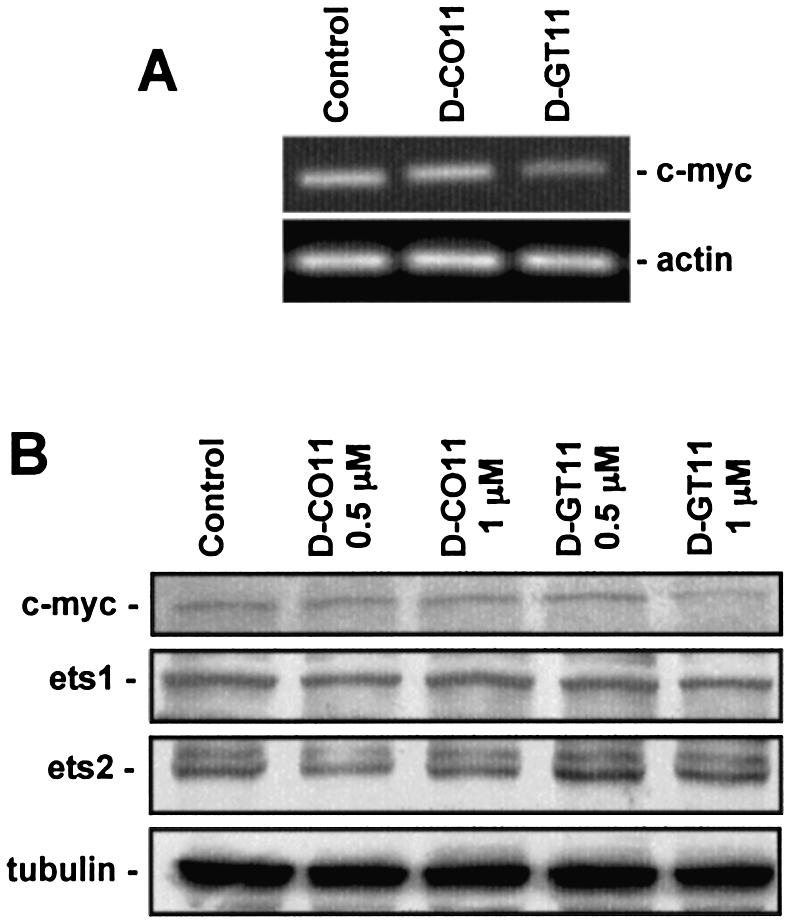
Triplex-forming oligonucleotides (TFO) that bind DNA in a sequence-specific manner might be used as selective repressors of gene expression and gene-targeted therapeutics. However, many factors, including instability of triple helical complexes in cells, limit the efficacy of this approach. In the present study, we tested whether covalent linkage of a TFO to daunomycin, which is a potent DNA-intercalating agent and anticancer drug, could increase stability of the triple helix and activity of the oligonucleotide in cells. The 11mer daunomycin-conjugated GT (dauno-GT11) TFO targeted a sequence upstream of the P2 promoter, a site known to be critical for transcription of the c-myc gene. Band-shift assays showed that the dauno-GT11 formed triplex DNA with enhanced stability compared to the unmodified TFO. Band shift and footprinting experiments demonstrated that binding of dauno-GT11 was highly sequence-specific with exclusive binding to the 11 bp target site in the c-myc promoter. The daunomycin-conjugated TFO inhibited transcription in vitro and reduced c-myc promoter activity in prostate and breast cancer cells. The daunomycin-conjugated TFO was taken up by cells with a distinctive intracellular distribution compared to free daunomycin. However, cationic lipid-mediated delivery was required for enhanced cellular uptake, nuclear localization and biological activity of the TFO in cells. Dauno-GT11 reduced transcription of the endogenous c-myc gene in cells, but did not affect expression of non-target genes, such as ets-1 and ets-2, which contained very similar target sequences in their promoters. Daunomycin-conjugated control oligonucleotides unable to form triplex DNA with the target sequence did not have any effect in these assays, indicating that daunomycin was not directly responsible for the activity of daunomycin-conjugated TFO. Thus, attachment of daunomycin resulted in increased triplex stability and biological activity of the 11mer GT-rich TFO without compromising its specificity. These results encourage further testing of this approach to develop novel antigene therapeutics.


Figures

Figure 1
Figure 1
Map of the c-myc gene and structure of the daunomycin-conjugated TFO. (A) The polypurine:polypyrimidine sequence near the P2 promoter of the c-myc gene and the oligonucleotides GT11 and CO11 are shown. The sites in the target duplex complementary to GT11 and CO11 are underlined. Lines under the sequence indicate the 23, 28 and 40 bp oligonucleotides used in band shift assays and the 11 bp target site. The m40bp is a mutated duplex in which four bases, shown in italics in the target sequence, had been changed to TCTC. Brackets above the gene map indicate the inserts cloned into the pMyc-1388 and pMyc-262, respectively. K, X and R indicate KpnI, XhoI and RsrII restriction sites. (B) Oligonucleotides were modified at the 3′ end with a propanediol tail and linked at the 5′ end to the ring D of the anthraquinone via a hexamethylene bridge. The three domains of the daunomycin-conjugated oligonucleotide are expected to bind by triplex formation in the major groove (oligonucleotide), minor groove binding (amino sugar) and intercalation at the duplex-triplex junction (anthraquinone), respectively.
Figure 1
Figure 1
Map of the c-myc gene and structure of the daunomycin-conjugated TFO. (A) The polypurine:polypyrimidine sequence near the P2 promoter of the c-myc gene and the oligonucleotides GT11 and CO11 are shown. The sites in the target duplex complementary to GT11 and CO11 are underlined. Lines under the sequence indicate the 23, 28 and 40 bp oligonucleotides used in band shift assays and the 11 bp target site. The m40bp is a mutated duplex in which four bases, shown in italics in the target sequence, had been changed to TCTC. Brackets above the gene map indicate the inserts cloned into the pMyc-1388 and pMyc-262, respectively. K, X and R indicate KpnI, XhoI and RsrII restriction sites. (B) Oligonucleotides were modified at the 3′ end with a propanediol tail and linked at the 5′ end to the ring D of the anthraquinone via a hexamethylene bridge. The three domains of the daunomycin-conjugated oligonucleotide are expected to bind by triplex formation in the major groove (oligonucleotide), minor groove binding (amino sugar) and intercalation at the duplex-triplex junction (anthraquinone), respectively.
Figure 2
Figure 2
Triplex DNA formation by non-conjugated and daunomycin-conjugated TFOs. Oligonucleotides corresponding to the pyrimidine-rich strands of the duplex targets were 5′ end-labeled with [γ-32P]ATP and annealed to the complementary oligonucleotides. Duplex DNA at a concentration of 1 nM was incubated for 18 h at 37°C with the indicated concentrations of non-conjugated GT11, dauno-GT11 or dauno-CO11. Gel electrophoresis was carried out under non-denaturating conditions at a gel temperature of 20 (A, B, D, E and F) or 10°C (C). Duplex targets were the 28 (A–D), 23 (E) and 40 bp (F) double-stranded oligonucleotides (see Fig. 1). Positions of duplex and triplex DNA are indicated.
Figure 3
Figure 3
Sequence-specific binding and inhibition of transcription initiation by the daunomycin-conjugated TFO. (A) Two 40 bp duplex oligonucleotides with either the wild-type (top) or a mutated (bottom) target sequence were incubated with dauno-GT11 or dauno-CO11 for 18 h at 37°C. Samples were then analyzed under non-denaturating conditions at a gel temperature of 37°C. The mutated site is shown in Figure 1. (B) A 32P-end-labeled 339 bp fragment of the c-myc promoter was incubated with or without dauno-GT11 and dauno-CO11 for 18 h at 37°C. DNA was then treated with DMS and piperidine to cleave unprotected guanines and samples were analyzed by gel electrophoresis under denaturating conditions. The position of the 11 nt target sequence is shown. (C) The p1388-Myc (top) and the pGL3control plasmid (bottom), which contained the SV40 promoter, were incubated without or with 2.5 µM of dauno-GT11 and dauno-CO11 for 18 h at 37°C. HeLa cell nuclear extract was added and samples were incubated for 1 h at 30°C. Following phenol/chloroform extraction and ethanol precipitation, samples were subjected to primer extension with a 32P-labeled primer. A radiolabeled internal control RNA (i.c.) was added to monitor sample recovery throughout the procedure. Samples were analyzed on a denaturating polyacrylamide gel. The positions of the transcripts generated from the c-myc and the SV40 promoter, respectively, are indicated.
Figure 4
Figure 4
Cellular uptake of free daunomycin and daunomycin-conjugated TFO. (A) MCF-7 cells were incubated with 0.2 and 1 µM of daunomycin or dauno-GT11. After 4 h, cells were washed, harvested and collected by centrifugation. Uptake was then determined by flow cytometry. Untreated cells (control) were used to determine background fluorescence levels. (B) MCF-7 cells were incubated with 1 µM of daunomycin (a and b) or dauno-GT11 (c and d) for 6 h and then examined by fluorescence (top) and phase-contrast (bottom) microscopy.
Figure 5
Figure 5
Uptake of daunomycin-conjugated TFO in the presence of cationic lipids. (A) DU145 cells were incubated with dauno-GT11 (0.5 µM) alone or combined with the transfection reagent DOTAP for 6 h. After 24 h, cells were harvested and analyzed by flow cytometry. Untreated cells (control) were used to determine background fluorescence levels. (B) DU145 cells incubated with dauno-GT11 complexed with DOTAP (a, b and c) or dauno-GT11 alone (d, e and f) were examined by fluorescence and phase-contrast microscopy. Fluorescence (top), phase-contrast (middle) and merged (bottom) images are shown for each treatment group.
Figure 6
Figure 6
Inhibition of c-myc promoter activity by daunomycin-conjugated TFO in prostate and breast cancer cells. Cells were transfected for 6 h either with p1388-Myc or p262-Myc reporter plasmid, a pRL control plasmid and the indicated concentrations of dauno-GT11 (empty bars) or dauno-CO11 (filled bars). Luciferase activity was measured after 24 h. Data are presented as percent of luciferase activity in TFO-treated cells compared to cells incubated with an equal concentration of control oligonucleotide. (A) DU145 cells transfected with p1388-Myc; (B) DU145 cells transfected with p262-Myc; (C) MCF-7 cells transfected with p262-Myc; (D) MDA-MB-231 cells transfected with p262-Myc.
Figure 7
Figure 7
Inhibition of c-myc promoter activity is dependent on the triplex-forming ability of daunomycin-conjugated TFO. (A) Structure of the daunomycin-conjugated control oligonucleotide dauno-GT11C. (B) Gel mobility shift assay with duplex DNA incubated without (control) or with dauno-GT11 (D-GT11), dauno-GT11C (D-GT11C) or non-conjugated GT11. (C) DU145 cells were transfected with p262-Myc reporter plasmid and pRL control vector in the presence of 1 µM of dauno-GT11 (empty bars), dauno-CO11 (filled bars) or dauno-GT11C (gray bars). Luciferase activity was measured as described in the legend to Figure 6.
Figure 8
Figure 8
Inhibition of endogenous c-myc gene expression by daunomycin-conjugated TFO. (A) DU145 cells were transfected for 6 h with 1 µM of dauno-GT11 or dauno-CO11 using DOTAP. After 24 h, total RNA was extracted from untreated (Control), control oligonucleotide- (D-CO11) and TFO- (D-GT11) treated cells. c-myc and β-actin RNA levels were determined by RT–PCR. (B) DU145 cells were transfected with 0.5 and 1 µM of dauno-GT11 or dauno-CO11 as indicated above. Western blot analysis was performed after 24 h to determine c-myc, ets-1, ets-2 and α-tubulin protein level.

References

    1. Chan P.P. and Glazer,P.M. (1997) Triplex DNA: fundamentals, advances and potential applications for gene therapy. Mol. Med., 75, 267–282. - PubMed
    1. Praseuth D., Guieysse,A.L. and Helene,C. (1999) Triple helix formation and the antigene strategy for sequence-specific control of gene expression. Biochim. Biophys. Acta, 1489, 181–206. - PubMed
    1. Casey B.P. and Glazer,P.M. (2001) Gene targeting via triple-helix formation. Prog. Nucleic Acid Res. Mol. Biol., 67, 163–192. - PubMed
    1. Winters T.A. (2000) Gene targeted agents: new opportunities for rational drug development. Curr. Opin. Mol. Ther., 2, 670–681. - PubMed
    1. Moser H.E. and Dervan,P.B. (1987) Sequence-specific cleavage of double helical DNA by triple helix formation. Science, 238, 645–650. - PubMed

Publication types

MeSH terms