. 2004 May;135(1):400-11.
doi: 10.1104/pp.103.037945. Epub 2004 Apr 30.

Structure and expression profile of the Arabidopsis PHO1 gene family indicates a broad role in inorganic phosphate homeostasis

Affiliations

Structure and expression profile of the Arabidopsis PHO1 gene family indicates a broad role in inorganic phosphate homeostasis

Yong Wang et al. Plant Physiol. 2004 May.

Abstract

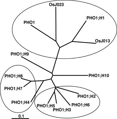
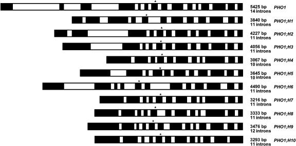
PHO1 has been recently identified as a protein involved in the loading of inorganic phosphate into the xylem of roots in Arabidopsis. The genome of Arabidopsis contains 11 members of the PHO1 gene family. The cDNAs of all PHO1 homologs have been cloned and sequenced. All proteins have the same topology and harbor a SPX tripartite domain in the N-terminal hydrophilic portion and an EXS domain in the C-terminal hydrophobic portion. The SPX and EXS domains have been identified in yeast (Saccharomyces cerevisiae) proteins involved in either phosphate transport or sensing or in sorting proteins to endomembranes. The Arabidopsis genome contains additional proteins of unknown function containing either a SPX or an EXS domain. Phylogenetic analysis indicated that the PHO1 family is subdivided into at least three clusters. Reverse transcription-PCR revealed a broad pattern of expression in leaves, roots, stems, and flowers for most genes, although two genes are expressed exclusively in flowers. Analysis of the activity of the promoter of all PHO1 homologs using promoter-beta-glucuronidase fusions revealed a predominant expression in the vascular tissues of roots, leaves, stems, or flowers. beta-Glucuronidase expression is also detected for several promoters in nonvascular tissue, including hydathodes, trichomes, root tip, root cortical/epidermal cells, and pollen grains. The expression pattern of PHO1 homologs indicates a likely role of the PHO1 proteins not only in the transfer of phosphate to the vascular cylinder of various tissues but also in the acquisition of phosphate into cells, such as pollen or root epidermal/cortical cells.


Figures

Figure 1.
Figure 1.
Identity/similarity matrix for the PHO1 family. Amino acid identity and similarity are indicated by the first and second number, respectively.
Figure 2.
Figure 2.
Alignment of the Arabidopsis PHO1 protein family. Alignment was done using the ClustalX program (Jeanmougin and Thompson, 1998; ftp://ftp-igbmc.u-strasbg.fr/pub/ClustalX/). The locations of the three SPX subdomains and of the EXS domain are indicated by a black bar over the sequence. The beginning of the hydrophobic C-terminal half of the proteins is indicated by an arrowhead. The protein sequence derived from two full-length rice cDNA clones J023079I02 and J013095I12 are indicated as OsJ023 and OsJ013, respectively.
Figure 3.
Figure 3.
Unrooted phylogenetic tree of the PHO1 family. The tree was made using the Neighbor-Joining method available on ClustalX (Jeanmougin and Thompson, 1998; ftp://ftp-igbmc.u-strasbg.fr/pub/ClustalX/).
Figure 4.
Figure 4.
Localization of the PHO1 gene family on the Arabidopsis chromosomes. Segmental duplications of the genome in areas containing PHO1 gene family members are indicated by links between or within chromosomes. Arrows indicate the relative orientation of the genes.
Figure 5.
Figure 5.
Intron/exon structure of the PHO1 gene family. Exons and introns are indicated by black and white boxes, respectively. The distances in base pairs between the start and stop codons on the genomic DNA are indicated on the right, along with the numbers of introns. Asterisks denote the location of the beginning of the hydrophobic region of the proteins.
Figure 6.
Figure 6.
Alignment of the SPX domain in Arabidopsis proteins. Alignment of the three SPX domains, referred as SPX-1 (a), SPX-2 (b), and SPX-3 (c), was done using the ClustalX program (Jeanmougin and Thompson, 1998; ftp://ftp-igbmc.u-strasbg.fr/pub/ClustalX/). The proteins included are the N. crassa (Nc) NUC2; the yeast (Sc) PHO81, PHO87, PHO90, PHO91, SYG1, and VCT3; the human (Hs) XPR1; the Arabidopsis (At) PHO1 family members PHO1, PHO1;H1, PHO1;H2, and PHO1;H3; as well as nine other Arabidopsis proteins of unknown functions. d, Unrooted phylogenetic tree of the all Arabidopsis proteins containing a SPX domain. The tree was made using the Neighbor-Joining method available on ClustalX.
Figure 7.
Figure 7.
Alignment of the EXS domain in Arabidopsis proteins. Alignment of EXS domain was done using the ClustalX program (Jeanmougin and Thompson, 1998; ftp://ftp-igbmc.u-strasbg.fr/pub/ClustalX/). The proteins included are the yeast (Sc) SYG1 and ERD1, the human (Hs) XPR1, the Arabidopsis (At) PHO1 family members PHO1, PHO1;H1, PHO1;H2, and PHO1;H3, as well as two other Arabidopsis proteins of unknown functions.
Figure 8.
Figure 8.
Expression analysis of the PHO1 gene family by RT-PCR. RNA was isolated from leaves (Le), stems (St), and flowers (Fl) from either wild-type Arabidopsis (WT + Pi) or the pho1 mutant (pho1) growing in soil. RNA was also harvested from roots (Ro) of plants grown in liquid nutrient media containing either 5 mm Pi (WT + Pi) or no Pi (−Pi). DNA fragment obtained by PCR performed on genomic DNA using the same oligonucleotides used for RT-PCR is shown in the last lane (DNA). This control ensures that the lower band obtained by RT-PCR cannot be derived from the amplification of genomic DNA that could be present in the RNA preparations. See “Material and Methods” for further details.
Figure 9.
Figure 9.
Localization of GUS activity in transgenic Arabidopsis expressing PHO1 gene family promoter-GUS fusion constructs. Promoter H3 activity in root vascular tissue (A), promoter H10 activity in root epidermal/cortical cells (B), promoter H7 activity in root tip (C), promoter H1 activity in leaf vascular tissue (D), promoter H7 activity in hydathodes (E), promoter H8 activity in trichomes (F–H), promoter H5 activity in petioles (I), promoter H10 activity in leaf blade (J), promoter H1 activity in stem vascular tissue (K), promoter H7 activity in stem (L), promoter H10 activity in pollen grain (M), promoter H9 activity in pollen grain (N), promoter H8 activity in stigma apex (O), promoter H9 activity in germinating pollen grain (P), promoter H1 activity in vascular tissue of sepals, petals, and filaments (Q), close-up of promoter H1 activity in filament vascular tissue (R), and promoter H6 activity in connective tissue of the anther (S) are shown.

References

    1. Auesukaree C, Homma T, Kaneko Y, Harashima S (2003) Transcriptional regulation of phosphate-responsive genes in low-affinity phosphate-transporter-defective mutants in Saccharomyces cerevisiae. Biochem Biophys Res Commun 306: 843–850 - PubMed
    1. Battini J-L, Rasko JEJ, Miller AD (1999) A human cell-surface receptor for xenotropic and polytropic murine leukemia viruses: possible role in G protein-coupled signal transduction. Proc Natl Acad Sci USA 96: 1385–1390 - PMC - PubMed
    1. Burleigh SH (2001) Relative quantitative RT-PCR to study the expression of plant nutrient transporters in arbuscular mycorrhizas. Plant Sci 160: 899–904 - PubMed
    1. Clough SJ, Bent AF (1998) Floral dip: a simplified method for Agrobacterium-mediated transformation of Arabidopsis thaliana. Plant J 6: 735–743 - PubMed
    1. Chomczynski P, Sacchi N (1987) Single-step method of RNA isolation by acid guanidium thiocyanate-phenol-chloroform extraction. Anal Biochem 162: 156–159 - PubMed

Publication types

MeSH terms