

The Human MitoChip: a high-throughput sequencing microarray for mitochondrial mutation detection
- PMID: 15123581
- PMCID: PMC479107
- DOI: 10.1101/gr.2228504
The Human MitoChip: a high-throughput sequencing microarray for mitochondrial mutation detection
Abstract
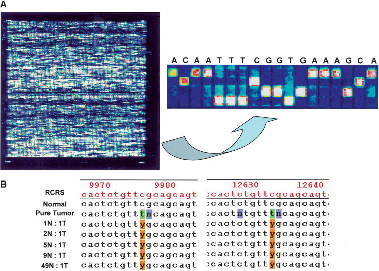
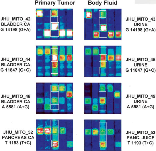
Somatic mitochondrial mutations are common in human cancers, and can be used as a tool for early detection of cancer. We have developed a mitochondrial Custom Reseq microarray as an array-based sequencing platform for rapid and high-throughput analysis of mitochondrial DNA. The MitoChip contains oligonucleotide probes synthesized using standard photolithography and solid-phase synthesis, and is able to sequence >29 kb of double-stranded DNA in a single assay. Both strands of the entire human mitochondrial coding sequence (15,451 bp) are arrayed on the MitoChip; both strands of an additional 12,935 bp (84% of coding DNA) are arrayed in duplicate. We used 300 ng of genomic DNA to amplify the mitochondrial coding sequence in three overlapping long PCR fragments. We then sequenced >2 million base pairs of mitochondrial DNA, and successfully assigned base calls at 96.0% of nucleotide positions. Replicate experiments demonstrated >99.99% reproducibility. In matched fluid samples (urine and pancreatic juice, respectively) obtained from five patients with bladder cancer and four with pancreatic cancer, the MitoChip detected at least one cancer-associated mitochondrial mutation in six (66%) of nine samples. The MitoChip is a high-throughput sequencing tool for the reliable identification of mitochondrial DNA mutations from primary tumors in clinical samples.
Figures
References
-
- Andrews, R.M., Kubacka, I., Chinnery, P.F., Lightowlers, R.N., Turnbull, D.M., and Howell, N. 1999. Reanalysis and revision of the Cambridge reference sequence for human mitochondrial DNA. Nat. Genet. 23: 147. - PubMed
-
- Bianchi, N.O., Bianchi, M.S., and Richard, S.M. 2001. Mitochondrial genome instability in human cancers. Mutat. Res. 488: 9-23. - PubMed
-
- Boland, C.R., Thibodeau, S.N., Hamilton, S.R., Sidransky, D., Eshleman, J.R., Burt, R.W., Meltzer, S.J., Rodriguez-Bigas, M.A., Fodde, R., Ranzani, G.N., et al. 1998. A National Cancer Institute workshop on microsatellite instability for cancer detection and familial predisposition: Development of international criteria for the determination of microsatellite instability in colorectal cancer. Cancer Res. 58: 5248-5257. - PubMed
-
- Chee, M., Yang, R., Hubbell, E., Berno, A., Huang, X.C., Stern, D., Winkler, J., Lockhart, D.J., Morris, M.S., and Fodor, S.P. 1996. Accessing genetic information with high-density DNA arrays. Science 274: 610-614. - PubMed
WEB SITE REFERENCES
-
- http://www.mitomap.org; 2003, MITOMAP: A Human Mitochondrial Genome Database. - PMC - PubMed
-
- http://engels.genetics.wisc.edu/amplify/; the Amplify 1.2 program.
Publication types
MeSH terms
Substances
Grants and funding
LinkOut - more resources
Full Text Sources
Other Literature Sources