. 2004 May 10;165(3):335-46.
doi: 10.1083/jcb.200403061. Epub 2004 May 3.

Degradative organelles containing mislocalized alpha-and beta-synuclein proliferate in presenilin-1 null neurons

Affiliations

Degradative organelles containing mislocalized alpha-and beta-synuclein proliferate in presenilin-1 null neurons

Christina A Wilson et al. J Cell Biol. .

Abstract

Presenilin-1 null mutation (PS1 -/-) in mice is associated with morphological alterations and defects in cleavage of transmembrane proteins. Here, we demonstrate that PS1 deficiency also leads to the formation of degradative vacuoles and to the aberrant translocation of presynaptic alpha- and beta-synuclein proteins to these organelles in the perikarya of primary neurons, concomitant with significant increases in the levels of both synucleins. Stimulation of autophagy in control neurons produced a similar mislocalization of synucleins as genetic ablation of PS1. These effects were not the result of the loss of PS1 gamma-secretase activity; however, dysregulation of calcium channels in PS1 -/- cells may be involved. Finally, colocalization of alpha-synuclein and degradative organelles was observed in brains from patients with the Lewy body variant of AD. Thus, aberrant accumulation of alpha- and beta-synuclein in degradative organelles are novel features of PS1 -/- neurons, and similar events may promote the formation of alpha-synuclein inclusions associated with neurodegenerative diseases.


Figures

Figure 1.
Figure 1.
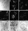
Enlarged lysosomal organelles are visible in PS1 −/− cells. (A) Differential contrast interference imaging of the cell surface of PS1 −/− fibroblasts demonstrated that a significant volume of the cells was occupied by large, spherical structures (arrows point to examples of enlarged vesicular structures). Lamp-2 immunolabeling of PS1 −/− (B) or PS1 +/? (C) fibroblasts demonstrated that these enlarged structures expressed lysosomal marker proteins and were present only within PS1 −/− cells. Similar accumulations of Lamp-2–positive organelles were detected in (D and E) PS1 −/− but not (F) PS1 +/? mouse primary neurons. Transmission EM analysis of PS1 −/− (G and H) or PS1 +/?. (I) Neurons demonstrated that PS1 −/− neurons contain large clusters of organelles. H is a higher magnification of the boxed area in G. *, denotes nucleus. Bars: (B–D and F) 10 μm; (E) 5 μm; (G and I) 1 μm; (H) 200 nm.
Figure 2.
Figure 2.
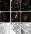
Perinuclear synuclein accumulations can be visualized in association with lysosomes in PS1 −/− neurons. (A) PS1 +/? or (B and C) PS1 −/− primary neuronal cultures were immunolabeled with antibodies to synaptophysin (green) and α-synuclein (A and B, red; Ab SNL-1) or synapsin I (green) and β-synuclein (C, red; Ab Syn 207). In PS1 −/− neurons, α- and β-synuclein are found in perinuclear locations that do not contain other presynaptic proteins (B and C, arrows). (D–F) Colocalization of lysosomal-associated membrane protein 2 (LAMP-2) with perinuclear synuclein accumulations in PS1 −/− neurons. (D) PS1 +/? or (E and F) PS1 −/− neurons cultured for 10 d were immunolabeled with antibodies to synuclein (Syn 202, red) and LAMP-2 (ABL93, green). (G–I) Immuno-EM of synuclein association with perinuclear multivesicular and autophagic organelles in PS1 −/− neurons. Synuclein is not found in association with lysosomes in PS1 +/? neurons (G, arrowhead) but is localized to enlarged lysosomal organelles in PS1 −/− neurons (H and I). *, denotes nucleus. Bars: (A–E) 10 μm; (F) 5 μm; (H) 500 nm; (G and I) 100 nm.
Figure 3.
Figure 3.
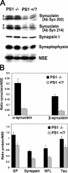
α- and β-synuclein are overexpressed in PS1/neurons. (A) Increased levels of both α- and β-synuclein in PS1 −/− lysates were demonstrated with the synuclein antibodies Syn 202 and Syn 214. Other synaptic proteins including synapsin I and synaptophysin were increased only slightly in PS1 −/− neurons. NSE was used as a loading control. (B) 125I-quantification of synuclein, synaptophysin, synapsin I, NFL, and tau levels relative to NSE. Error bars represent SD.
Figure 4.
Figure 4.
Expression of human wild-type or FAD mutant PS1 eliminated synuclein perinuclear accumulations and overexpression. (A–D) Immunolabeling of neuronal cultures with antibodies to synapsin I (green) and α- and β-synuclein (red, Syn 202). Neuronal cultures were generated from mice (A) PS1 −/−, (B) PS1 +/?, (C) PS1 −/− rescued with wild-type human PS, or (D) PS1 −/− rescued with PS1 containing FAD mutant A246E. Perinuclear synuclein accumulations appeared only in PS1 −/− neurons (A, arrow). *, denotes nucleus. Bar, 10 μm. (E) Western blots of lysates from 10-d-old cultures. The mouse PS1 carboxy-terminal fragment was present only in PS1 +/? cultures, and the larger human fragment was present only in the two rescue lines. α- and β-synuclein levels were increased solely in the PS1 −/− cultures. NSE was used as a loading control.
Figure 5.
Figure 5.
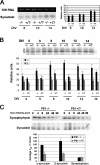
Examination of synuclein synthesis and degradation in PS1/neurons. (A) Northern blot and PhosphorImager quantification of synuclein mRNA levels in PS1 −/− and PS1 +/? neurons after multiple DIV. α- and β-synuclein appeared as one band on the Northern blot. There were no differences in the levels of synuclein mRNA. (B) Immunoblot and quantification of synuclein translation. Translation of α-synuclein, but not β-synuclein, was significantly increased in PS1 −/− neurons. (C) Pulse chase and quantification of synuclein and synaptophysin turnover. The average half-life of both α- and β-synuclein was increased in PS1 −/− neurons, whereas that of synaptophysin was unchanged between the two cell types. Error bars represent SD.
Figure 6.
Figure 6.
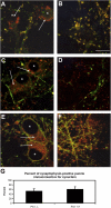
Axonal transport is not impaired in PS1 −/− neurons. The localization of proteins redistributed by the fast rate component of axonal transport (A and B, synaptophysin, green) as well as the slow rate component (C and D, NFL, green; E and F, β-tubulin, green) was compared with that of α- and β-synuclein (red, Syn 202) in (A, C, and E) PS1 −/− neurons and (B, D, and F) PS1 +/? neurons. None of the proteins examined appeared localized to the perinuclear synuclein accumulations. *, denotes nucleus. Bar, 5 μm. Arrows illustrate perinuclear synuclein accumulation. (G) The percentage of synaptophysin-positive putative synapses colabeled with synuclein immunoreactivity. Error bars represent SD.
Figure 7.
Figure 7.
Induction of autophagy with staurosporine stimulates the association of synucleins with perinuclear lysosomal organelles. PS1 +/? neurons were treated with 100 nM staurosporine for 48 h to induce autophagy. Immunolabeling of (A–C) untreated and (D–F) treated neurons with antibodies to Lamp-2 (A and D, green) and synuclein (B and E, Syn 202, red). The overlap is shown in C and F. Nuclei were counterstained with DAPI (blue). Bar, 5 μm.
Figure 8.
Figure 8.
Loss of PS1 γ-secretase activity does not result in the formation of enlarged perinuclear organelles. PS1 +/? neurons were treated for 7 d with the γ-secretase inhibitors L-685,458 (1 μM), DFK-167 (50 μM), III-31C (5 μM), 2-Napthyl-VF-CHO (20 μM), or LiCl (5 mM), and analyzed by light microscopy for Lamp-2 and synuclein immunoreactivity. None of these compounds increased the percentage of neurons containing Lamp-2–positive enlarged perinuclear organelles, nor did they induce the localization of synuclein proteins to this site. 100 nM staurosporine treatment for 48 h was included as a positive control. Asterisks indicate that the results are statistically significant, P < 0.005. Error bars represent SD.
Figure 9.
Figure 9.
Inhibition of capacitative calcium channels eliminates the perinuclear distribution of synucleins in PS1/neurons. PS1 −/− and PS1 +/? neurons cultured for 10 d in vitro were treated with SKF 96365 or thapsigargin, or incubated in low Ca2+ medium for 48 h. Representative images of (A) untreated PS1 +/? neurons, (B) untreated PS1 −/−neurons, or (C) PS1 −/− neurons treated with 500 nM SKF 96365 are shown. Nuclei are counterstained with DAPI (blue). Bar, 10 μm. (D) Quantification of the percentage of neurons displaying perinuclear synuclein immunoreactivity. Error bars represent SD.
Figure 10.
Figure 10.
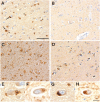
Perinuclear α-synuclein deposits colocalize with Lamp-immunoreactivity in dentate hilar neurons from patients with LBVAD. Hippocampal tissue from (A) LBVAD patients immunolabeled with Syn 303 revealed perinuclear synuclein immunoreactivity in neuronal cell bodies (arrows) and neurites (*) as well as near the nuclei of nonneuronal cells (arrowheads), which are not seen in tissue from (B) nondiseased controls. Similarly, Lamp-2 immunoreactivity is increased in (C) LBVAD dentate gyrus relative to (D) controls. A comparison of synuclein immunoreactivity (E and G, Syn 303) with Lamp-2 immunoreactivity in adjacent sections (F and H, Lamp-2 C20) demonstrated similar localization of the two proteins in cell bodies. Bars: (A–D) 30 μm; (E–H) 10 μm.

Similar articles

Cited by

References

    1. Adamec, E., P.S. Mohan, A.M. Cataldo, A.N. Srinivasan, J.P. Vonsattel, and R.A. Nixon. 2000. Upregulation of the lysosomal system in experimental models of neuronal injury: Implications for Alzheimer's disease. Neuroscience. 100:663–675. - PubMed
    1. Advani, R.J., B. Yang, R. Prekeris, K.C. Lee, J. Klumperman, and R.H. Scheller. 1999. VAMP-7 mediates vesicular transport from endosomes to lysosomes. J. Cell Biol. 146:765–775. - PMC - PubMed
    1. Anglade, P., S. Vyas, F. Javoy-Agid, M.T. Herrero, P.P. Michel, J. Marquez, A. Mouatt-Prigent, M. Ruberg, E.C. Hirsch, and Y. Agid. 1997. Apoptosis and autophagy in nigral neurons of patients with Parkinson's disease. Histol. Histopathol. 12:25–31. - PubMed
    1. Annaert, W.G., C. Esselens, V. Baert, C. Boeve, G. Snellings, P. Cupers, K. Craessaerts, and B. De Strooper. 2001. Interaction with telencephalin and the amyloid precursor protein predicts a ring structure for presenilins. Neuron. 32:579–589. - PubMed
    1. Bembi, B., M.S. Zambito, E. Sidransky, G. Ciana, M. Carrozzi, C. Martini, M. Giolis, M.G. Pittis, and L. Capus. 2003. Gaucher's disease with Parkinson's disease: Clinical and pathogical aspects. Neurology. 61:99–101. - PubMed

Publication types

MeSH terms