. 2004 May 3;199(9):1245-54.
doi: 10.1084/jem.20031989.

2B4 acts as a non-major histocompatibility complex binding inhibitory receptor on mouse natural killer cells

Affiliations

2B4 acts as a non-major histocompatibility complex binding inhibitory receptor on mouse natural killer cells

Kyung-Mi Lee et al. J Exp Med. .

Abstract

Natural killer (NK) cells are critical in the immune response to tumor cells, virally infected cells, and bone marrow allografts. 2B4 (CD244) is expressed on all NK cells and the ligand for 2B4, CD48, is expressed on hematopoietic cells. Cross-linking 2B4 on NK cells with anti-2B4 monoclonal antibody leads to NK cell activation in vitro. Therefore, 2B4 is considered to be an activating receptor. Surprisingly, we have found, using antibody-blocking and 2B4-deficient NK cells, that NK lysis of CD48(+) tumor and allogeneic targets is inhibited by 2B4 ligation. Interferon gamma production by NK cells is also inhibited. Using a peritoneal tumor clearance assay, it was found that 2B4(-/-) mice have increased clearance of CD48(+) tumor cells in vivo. Retroviral transduction of 2B4 was sufficient to restore inhibition in 2B4(-/-) primary NK cells. It was found that although mature NK cells express SH2D1A, in vitro-derived NK cells do not. However, both populations are inhibited by 2B4 ligation. This indicates that 2B4 inhibitory signaling occurs regardless of the presence of SH2D1A. These findings reveal a novel role for 2B4 as a non-major histocompatibility complex binding negative regulator of NK cells.


Figures

Figure 1.
Figure 1.
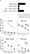
Ligation of 2B4 by CD48 expressed on tumor cells inhibits NK cytotoxicity. (A) CD48 and CD48+ RMA-S cells were stained with anti-CD48 (open histogram) or isotype control (shaded histogram). (B) WT LAK cells were tested in a 4-h chromium release assay against CD48 or CD48+ RMA-S cells at the indicated E/T ratios. (C) WT LAK cells were pretreated with anti-FcR and either anti-2B4 or isotype control and tested against CD48 or CD48+ RMA-S. (D) WT LAK cells were tested against CD48+ RMA-S. NK cells were pretreated with anti-FcR and targets were pretreated with or without anti-CD48. (E) CD48 RMA-S cells were transduced with CD48 or control empty vector and tested as targets of WT NK cells. (F) WT LAK cells were pretreated with anti-2B4 or isotype control antibody and tested for killing of CD48 P815 or P815 transfected with CD48. (G) WT and 2B4−/− LAK cells were tested against CD48 and CD48+ RMA-S targets.
Figure 2.
Figure 2.
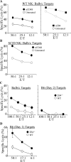
Ligation of 2B4 by CD48 expressed on allogeneic and syngeneic targets inhibits NK cytotoxicity. B6 LAK cells were tested in a cytotoxicity assay against Con A–stimulated splenic cells at the indicated E/T ratios. (A) On the left, WT LAK cells were tested against BALB/c targets either pretreated with anti-CD48 or not treated. On the right, WT LAK cells pretreated with anti-2B4 or not treated were tested against BALB/c targets. (B) SCID LAK cells were tested against BALB/c targets either pretreated with anti-CD48 or untreated. (C) WT and 2B4−/− LAK cells were assayed against BALB/c or B6 Con A blasts. (D) WT and 2B4−/− LAK cells were tested against B cell–depleted B6 splenocytes stimulated with Con A for 1 d only.
Figure 3.
Figure 3.
Ligation of 2B4 on in vitro–derived Ly49 NK cells by CD48 inhibits cytotoxicity. (A) WT in vitro–derived NK cells were tested in a 4-h chromium release assay against CD48 and CD48+ RMA-S in the presence of anti-CD48, isotype control, or no antibody at a 50:1 E/T ratio. (B) WT and 2B4−/− in vitro–derived NK cells were tested against CD48 or CD48+ RMA-S at the indicated E/T ratios. (C) WT and 2B4−/− in vitro–derived NK cells were tested against Con A–stimulated BALB/c or B6 splenic cells at the indicated E/T ratios.
Figure 4.
Figure 4.
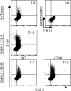
CD48 on target cells inhibits NK cell IFN-γ production. WT LAK cells pretreated with anti-FcR were incubated at a 1:1 ratio with CD48 or CD48+ RMA-S cells for 6 h. CD48+ RMA-S were pretreated with anti-CD48 or isotype control. NK cells incubated alone were used as controls. Cells were gated on the basis of forward and size scatter to exclude RMA-S and were gated on CD3 NK1.1+ cells. Numbers indicate the percent of NK cells that were IFN-γ+.
Figure 5.
Figure 5.
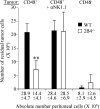
2B4 engagement inhibits NK rejection of tumor cells in vivo. WT and 2B4−/− mice were injected i.p. with CFSE-labeled tumor cells. The number of tumor cells recovered from the peritoneum after 3 d (depicted on y axis) was calculated on the basis of absolute number of peritoneal exudate cells (listed on x axis) multiplied by the percentage that were tumor cells as determined by flow cytometry. NK-depleted mice were treated with 100 μg anti-NK1.1 on days −2 and 0. Five mice per group were analyzed per experiment. Data are representative of three independent experiments. **, Student's t test for WT versus 2B4−/−; P = 0.0004.
Figure 6.
Figure 6.
Retroviral transduction of 2B4 long into 2B4−/− NK cells restores inhibition. 2B4−/− LAK cells were retrovirally transduced with a vector expressing 2B4 short, 2B4 long, or empty vector control. (A) 24 h after transduction, cells were analyzed for 2B4 expression (B) and for live cells by forward and side scatter. Numbers indicate the percent of cells in the live gate. (C) Transduced cells were tested for killing of CD48+ RMA-S. (D) In a separate experiment, transduced cells were tested for lysis of CD48 or CD48+ RMA-S. Data are representative of at least three independent experiments.
Figure 7.
Figure 7.
SH2D1A is differentially expressed in LAK and in vitro–derived NK cells. Total RNA was isolated from day 5 and day 11 in vitro–derived NK cells, LAK cells, and thymocytes as a control. RT-PCR was performed for (A) SH2D1A and (B) EAT-2 mRNA in the presence and absence of RT. Primers specific for GAPDH were used as a control.

References

    1. Biron, C.A., K.B. Nguyen, G.C. Pien, L.P. Cousens, and T.P. Salazar-Mather. 1999. Natural killer cells in antiviral defense: function and regulation by innate cytokines. Annu. Rev. Immunol. 17:189–220. - PubMed
    1. Lanier, L.L. 1995. The role of natural killer cells in transplantation. Curr. Opin. Immunol. 7:626–631. - PubMed
    1. Lanier, L.L. 1998. NK cell receptors. Annu. Rev. Immunol. 16:359–393. - PubMed
    1. McQueen, K.L., and P. Parham. 2002. Variable receptors controlling activation and inhibition of NK cells. Curr. Opin. Immunol. 14:615–621. - PubMed
    1. Lanier, L.L. 2001. On guard–activating NK cell receptors. Nat. Immunol. 2:23–27. - PubMed

Publication types

MeSH terms