Comparative Study
. 2004 May 3;199(9):1255-64.
doi: 10.1084/jem.20031835.

The cell surface receptor SLAM controls T cell and macrophage functions

Affiliations
Comparative Study

The cell surface receptor SLAM controls T cell and macrophage functions

Ninghai Wang et al. J Exp Med. .

Abstract

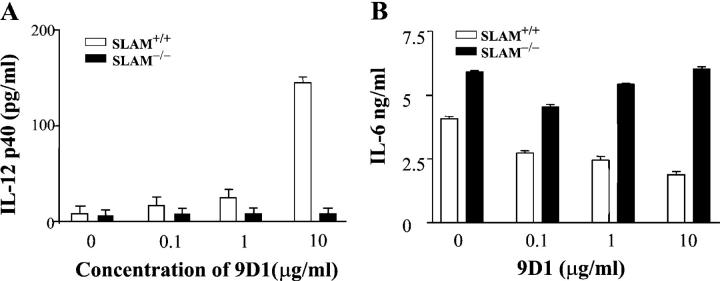
Signaling lymphocyte activation molecule (SLAM), a glycoprotein expressed on activated lymphocytes and antigen-presenting cells, has been shown to be a coregulator of antigen-driven T cell responses and is one of the two receptors for measles virus. Here we show that T cell receptor-induced interleukin (IL)-4 secretion by SLAM(-/-) CD4(+) cells is down-regulated, whereas interferon gamma production by CD4(+) T cells is only slightly up-regulated. Although SLAM controls production of IL-12, tumor necrosis factor, and nitric oxide in response to lipopolysaccharide (LPS) by macrophages, SLAM does not regulate phagocytosis and responses to peptidoglycan or CpG. Thus, SLAM acts as a coreceptor that regulates signals transduced by the major LPS receptor Toll-like receptor 4 on the surface of mouse macrophages. A defective macrophage function resulted in an inability of SLAM(-/-) C57Bl/6 mice to remove the parasite Leishmania major. We conclude that the coreceptor SLAM plays a central role at the interface of acquired and innate immune responses.


Figures

Figure 1.
Figure 1.
Targeted disruption of the mouse SLAM gene. (A) The targeting vector. Configuration of the WT SLAM genomic locus, the targeting vector, and the chromosomal locus after homologous recombination with the targeting vector is shown. The second and third exons of the SLAM gene, IgV and IgC, encoding the complete ectodomain of SLAM, were replaced with the neomycin resistance gene (Neo). An 8.2- and a 3.8-kb genomic DNA fragment flank the neomycin resistance gene. The locations of the two probes used in the Southern blotting analysis are shown. SP, signal peptide; TM, transmembrane region; CP1, CP2, and CP3, cytoplasmic domains; S, SacII; K, KpnI. (B) Southern blot analysis. Screening for homologous recombination events by Southern blotting of tail DNA samples used the 3′ probe in conjunction with a KpnI (K) digest. A 13-kb band is detected in WT and heterozygote mice. An 8.8-kb band, which results from a new KpnI site in the targeting vector, is detected in heterozygotes (+/−) and homozygotes (−/−). (C) RT-PCR analysis. RT-PCR products were generated with RNA from the thymus or spleen of SLAM+/+ or SLAM−/− mice. Although an SP plus CP3 primer pair detects a 1.1-kb RT-PCR product in SLAM+/+ thymocytes (T) or splenocytes (S), no RT-PCR product is detected in the thymus or spleen of SLAM−/− mice. (D) Absence of SLAM surface expression in SLAM-deficient thymocytes. SLAM−/− or SLAM+/+ thymocytes were stained with a monoclonal rat anti–mouse SLAM antibody (9D1) and analyzed by flow cytometry.
Figure 2.
Figure 2.
Deviation of cytokine production by CD4+ and CD8+ SLAM−/− T cells. (A) IL-4 secretion by CD4+ cells purified from SLAM−/− or SLAM+/+ C57BL/6 mice. SLAM−/− or SLAM+/+ C57BL/6 CD4+ cells were purified using negative selection columns. In the primary stimulation (−1°−) CD4+ cells were stimulated with 5 μg/ml anti-CD3ɛ antibody (2C11)–coated plates and 5 μg/ml soluble anti-CD28, or with a combination of 10 ng/ml PMA and 1 μM ionomycin in the presence of 10 ng/ml recombinant IL-2 (BD Biosciences; P/I) for 72 h. IL-4 in the cell culture supernatants was determined by ELISA. (B) IFN-γ secretion by CD4+ cells purified from SLAM−/− or SLAM+/+ C57BL/6 mice. SLAM−/− or SLAM+/+ C57BL/6 CD4+ cells were purified using negative selection columns. CD4+ cells were stimulated as described in A and IFN-γ in the cell culture supernatants was determined by ELISA. (C) Impaired IL-4 secretion by SLAM−/− × DO11.10 CD4+ and SAP−/− × DO11.10 CD4+ cells upon stimulation with APCs and OVA peptide. IL-4 secretion after primary (−1°−) and secondary (−2°−) stimulation of CD4+ T cells isolated from SLAM−/− × DO11.10, SAP−/− × DO11.10, or DO11.10 TCR transgenic mice were determined. Equal numbers of purified KJ-126+/CD4+ T cells isolated from SLAM−/− × DO11.10, SAP−/− × DO11.10, or DO11.10 TCR transgenic BALB/c mice (2 × 10⁵ cells/well) were stimulated for 3 d with APCs (2 × 10⁶) pulsed with 1.0 μ of the OVA peptide OVA 323–339. Secondary stimulations were performed for 24 h under the same conditions. IL-4 secretion was determined at day 3 of the primary stimulation and 24 h after the secondary stimulation by ELISA. (D) IFN-γ secretion by SLAM−/− × DO11.10 CD4+ and SAP−/− × DO11.10 CD4+ cells upon stimulation with APCs and OVA peptide. Cells described in C were analyzed for IFN-γ by ELISA after primary (−1°−) and secondary (−2°−) stimulation. (E) Transcription of IL-4 and IFN-γ in NKT cells after anti-CD3 administration. RT-PCR products of IL-4, IFN-γ, and GAPDH were generated with spleen RNA samples isolated from anti-CD3 or PBS-injected mice. SLAM−/− C67BL/6 and WT C67BL/6 littermates as well as SLAM−/− BALB/c and WT BALB/c were used. The RT-PCR products were analyzed on a 1% agarose gel and visualized by UV light.
Figure 3.
Figure 3.
Altered IL-12, TNF-α, IL-6, and NO production by peritoneal macrophages from SLAM−/− C57BL/6 mice. (A) IL-12 production by SLAM−/− C57BL/6 or WT SLAM+/+ C57BL/6 peritoneal macrophages. Resting peritoneal macrophages from 12 SLAM−/− C57BL/6 or WT SLAM+/+ C57BL/6 mice were stimulated with 200 U/ml IFN-γ, 2 ng/ml LPS, or with IFN-γ plus LPS for 24 h. IL-12p70 was determined by ELISA as described in Materials and Methods. M, cells incubated in medium only. (B) TNF-α production by SLAM−/− C57BL/6 or WT SLAM+/+ C57BL/6 peritoneal macrophages. Resting peritoneal macrophages from SLAM−/− C57BL/6 or WT SLAM+/+ C57BL/6 mice were stimulated as described in A. TNF-α was determined by ELISA in the same culture supernatants as described in Materials and Methods. M, cells incubated in medium only. (C) IL-6 production by SLAM−/− C57BL/6 or WT SLAM+/+ C57BL/6 peritoneal macrophages. Resting peritoneal macrophages from SLAM−/− C57BL/6 or WT SLAM+/+ C57BL/6 mice were stimulated as described in A. IL-6 was determined by ELISA in the same culture supernatants as described in Materials and Methods. M, cells incubated in medium only. (D) NO production by SLAM−/− C57BL/6 or WT SLAM+/+ C57BL/6 peritoneal macrophages. Resting peritoneal macrophages from SLAM−/− C57BL/6 or WT SLAM+/+ C57BL/6 mice were stimulated as described in A. NO was determined in the same culture supernatants as described in Materials and Methods. M, cells incubated in medium only. (E) IL-12p40 production is impaired in thioglycollate-induced peritoneal macrophages from SLAM−/− C57BL/6 mice, but not from SAP−/− C57BL/6 mice. Thioglycollate-elicited macrophages were isolated from the peritoneal cavity of SLAM−/−, SAP−/−, or WT SLAM+/+ C57BL/6 mice. 2 × 10⁶ macrophages were activated with increasing amounts of LPS (at 1, 10, or 100 ng/ml). Supernatants were collected after 24 h and IL-12p40 was determined by ELISA. (F) IL-12p40 production by macrophages in response to CpG and PGN. Thioglycollate-elicited peritoneal SLAM−/− or SLAM+/+ BALB/c macrophages (106 per well) were treated with 10 μM CpG or 25 μg/ml peptidoglycan for 24 h. Supernatants were removed and IL-12p40 levels were determined by ELISA. (G) TNF-α production by macrophages in response to CpG and PGN. Thioglycollate-elicited peritoneal SLAM−/− or SLAM+/+ BALB/c macrophages (106 per well) were treated with 10 μM CpG or 25 μg/ml peptidoglycan for 24 h. Supernatants were used to determine IL-12p40 levels by ELISA. (H) Antigen presentation by macrophages. 2 × 10⁵ thioglycollate-elicited peritoneal SLAM−/− or SLAM+/+ BALB/c macrophages were pulsed with 0.01–1 μg/ml of the OVA peptide OVA 323–339. Next, antigen-pulsed macrophages were incubated for 3 d with 105 KJ126+ CD4+ T cells isolated from DO11.10 TCR transgenic mice. To determine DNA synthesis, cultures were pulsed with [3H]thymidine in the final 18 h. (I) Phagocytosis of F18 E. coli by macrophages. Thioglycollate-elicited peritoneal SLAM−/− or SLAM+/+ BALB/c macrophages were incubated with E. coli F18 bacteria. After 1 h, cells were lysed and phagocytosis rates were obtained by counting colonies after 24 h of incubation. CFU, colony-forming units.
Figure 4.
Figure 4.
Monoclonal anti-SLAM antibody regulates IL-6 and IL-12 production by WT macrophages. IL-12 and IL-6 production by SLAM−/− or SLAM+/+ C57BL/6 thioglycollate-elicited peritoneal macrophages was determined in response to the anti-SLAM antibody 9D1. 106 peritoneal macrophages from SLAM−/− or SLAM+/+ C57BL/6 mice were treated for 24 h with 9D1 at the concentrations indicated. To each well, a rat anti–mouse IgG1 isotype control antibody was added (BD Biosciences) in a concentration range from 10 to 0 μg/ml, resulting in a final concentration of 10 μg/ml Ig in each experimental point. Supernatants were collected and analyzed for IL-12 p40 (A) or IL-6 (B) by ELISA.
Figure 5.
Figure 5.
SLAM−/− C57BL/6 mice are not resistant to infection with L. major. (A) Response to L. major infection by SLAM−/− BALB /c mice. SLAM−/− or SLAM+/+ BALB/c mice were infected in one footpad with 106 L. major stationary phase promastigotes. The size of footpad lesions of 10 infected mice of each strain was measured until 9 wk after infection and the change in footpad depth was plotted against time (in weeks). (B) Response to L. major infection by SLAM−/− C57BL/6 mice. SLAM−/− or SLAM+/+ C57BL/6 mice were infected with L. major as described in A. 30 mice were used in 3 experiments. A representative independent experiment with 12 mice is shown. ▴, SLAM−/− mice; ▪, SLAM+/+ mice. (C) Response to L. major infection by SAP−/− C57BL/6 mice. 10 SAP−/− or WT (SAP+/+) C57Bl/6 mice were infected with L. major as described in A. ▴, SAP−/− mice; ▪, WT SAP+/+ mice.

References

    1. Cocks, B.G., C.C. Chang, J.M. Carballido, H. Yssel, J.E. de Vries, and G. Aversa. 1995. A novel receptor involved in T-cell activation. Nature. 376:260–263. - PubMed
    1. Engel, P., M.J. Eck, and C. Terhorst. 2003. The SAP and SLAM families in immune responses and X-linked lymphoproliferative disease. Nat. Rev. Immunol. 3:813–821. - PubMed
    1. Tatsuo, H., N. Ono, K. Tanaka, and Y. Yanagi. 2000. SLAM (CDw150) is a cellular receptor for measles virus. Nature. 406:893–897. - PubMed
    1. Sayos, J., C. Wu, M. Morra, N. Wang, X. Zhang, D. Allen, S. van Schaik, L. Notarangelo, R. Geha, M.G. Roncarolo, et al. 1998. The X-linked lymphoproliferative-disease gene product SAP regulates signals induced through the co-receptor SLAM. Nature. 395:462–469. - PubMed
    1. Latour, S., G. Gish, C.D. Helgason, R.K. Humphries, T. Pawson, and A. Veillette. 2001. Regulation of SLAM-mediated signal transduction by SAP, the X-linked lymphoproliferative gene product. Nat. Immunol. 2:681–690. - PubMed

Publication types

MeSH terms