. 2004 May 11;101(19):7445-50.
doi: 10.1073/pnas.0401883101. Epub 2004 May 3.

The soluble ectodomain of herpes simplex virus gD contains a membrane-proximal pro-fusion domain and suffices to mediate virus entry

Affiliations

The soluble ectodomain of herpes simplex virus gD contains a membrane-proximal pro-fusion domain and suffices to mediate virus entry

Francesca Cocchi et al. Proc Natl Acad Sci U S A. .

Abstract

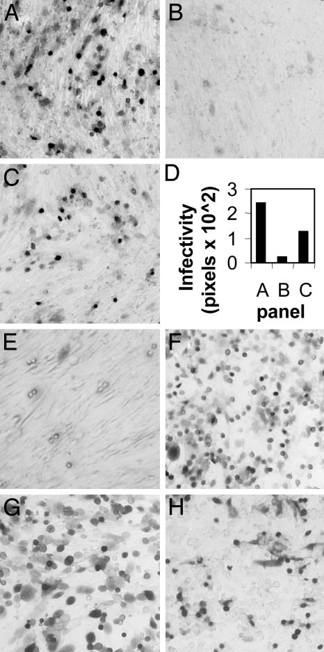
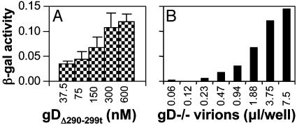
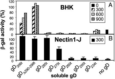
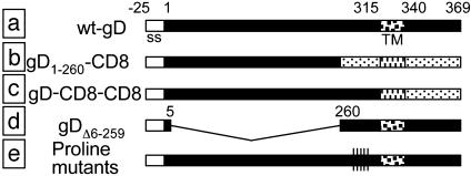
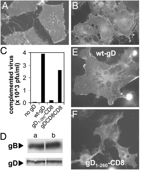
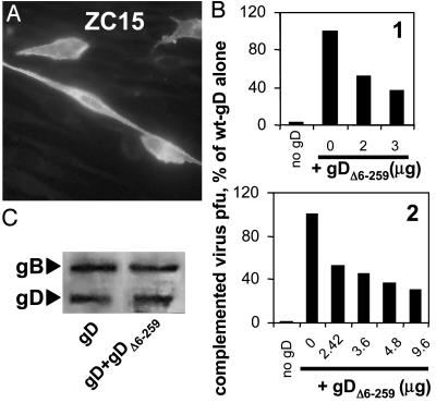
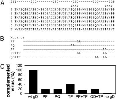
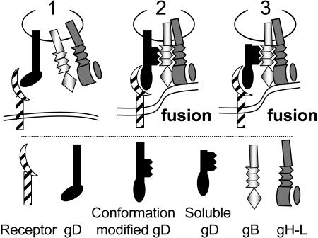
Entry of herpes simplex virus (HSV) 1 into cells requires the interaction of HSV gD with herpesvirus entry mediator or nectin1 receptors, and fusion with cell membrane mediated by the fusion glycoproteins gB, gH, and gL. We report that the gD ectodomain in soluble form (amino acids 1-305) was sufficient to rescue the infectivity of a gD-null HSV mutant, indicating that gD does not need to be anchored to the virion envelope to mediate entry. Entry mediated by soluble gD required, in addition to the receptor-binding sites contained within residues 1-250, a discrete downstream portion (amino acids 261-305), located proximal to the transmembrane segment in full-length gD. We named it as profusion domain. The pro-fusion domain was required for entry mediated by virion-bound gD, because its substitution with the corresponding region of CD8 failed to complement the infectivity of gD(-/+) HSV. Furthermore, a receptor-negative gD (gD(Delta6-259)) inhibited virus infectivity when coexpressed with wild-type gD; i.e., it acted as a dominant-negative gD mutant. The pro-fusion domain is proline-rich, which is characteristic of regions involved in protein-protein interactions. P291L-P292A substitutions diminished the gD capacity to complement gD(-/+) HSV infectivity. We propose that gD forms a tripartite complex with its receptor and, by way of the proline-rich pro-fusion domain, with the fusion glycoproteins, or with one of them. The tripartite complex would serve to recruit/activate the fusion glycoproteins and bring them from a fusion-inactive to a fusion-active state, such that they execute fusion of the virion envelope with cell membrane.


Figures

Fig. 1.
Fig. 1.
Rescue of gD-/- HSV infectivity by gDΔ290-299t (300 nM). (A and B) Cells infected with gD-/- HSV (3 μl) in the presence of gDΔ290-299t (A) or fetuin (B). Virions were mixed with glycoprotein during 150 min adsorption at 37°C. (C) The same amount of gD-/- HSV used in A was preincubated with gDΔ290-299t for 1 h at 4°C, and was then overlaid on BHK cells for 150 min. (D) Quantification of the results. Digital micrographs were taken with a 1.5× objective. The green areas (corresponding to infected cells) were quantified by the histogram program of Photoshop, and expressed in pixels. (E) A total of 200 μl of gD-/- virions were preincubated with 300 nM gDΔ290-299t for 1 h at 4°C, and were pelleted by centrifugation at 215,000 × g for 40 min. Virions were resuspended in 150 μl of DMEM and 100 μl were used to infect BHK cells. A-E, BHK cells. COS (F), nectin1-J (G), and J cells expressing HVEM (H) were infected as in A. J cells expressing HVEM were not cloned. All cells stained with 5-bromo-4-chloro-3-indolyl β-d-galactoside.
Fig. 2.
Fig. 2.
Rescue of gD-/- HSV infectivity by gDΔ290-299t is dose-dependent with respect to gD (A) and to virus (B). (A) Triplicate aliquots (3 μl) of gD-/- HSV were mixed with the indicated concentrations of gDΔ290-299t and were overlaid on BHK cells. (B) Increasing amounts of gD-/- virions were mixed with 300 nM gDΔ290-299t and were overlaid on BHK cells. β-galactosidase activity was measured at 16 h after infection. Bars denote ± SE.
Fig. 3.
Fig. 3.
Effect of different gDs, truncated at different residues in the ectodomain, on rescue of gD-/- HSV infectivity. BHK (A), or nectin1-J (B) cells were exposed to replicate aliquots of gD-/- HSV mixed with the indicated soluble gDs. gD was present at the indicated nM concentrations in A, and at 300 nM in B. The value obtained with 300 nM gDΔ290-299t is 100%. Each column represents the average of duplicates in A, and of triplicates in B. Bars denote ± SE.
Fig. 4.
Fig. 4.
Schematic representation of the gD constructs used in this study. (a) wt-gD. Empty bar, signal sequence (ss) cleaved in mature gD. The TM coordinates are marked. (b) gD1-260-CD8 chimera. (c) gD-CD8-CD8 from ref. . (d) Receptor-negative gDΔ6-259, the region between residues +6 and +259 was collapsed. (e) Proline mutants.
Fig. 5.
Fig. 5.
(A)gD1-260-CD8 expression in COS cells, detected by immunofluorescence with mAb H170. (B) Binding of a soluble nectin1 chimera made of nectin1 ectodomain fused to the constant fragment of human Ig (ref. ; 1.6 ng/μl) to paraformaldehyde-fixed gD1-260-CD8-expressing COS cells, detected by means of FITC-conjugated anti-human IgG Ab. (C) Infectivity complementation by gD1-260-CD8, gD-CD8-CD8, wt-gD, or no gD. Four h after transfection, BHK cells were infected with gD-/+ HSV (3 pfu per cell), and were then rinsed with pH 3 citrate buffer. Complemented virus was titrated at 24 h in R6 cells. (D) Quantification of gD, and of gB as control, present in complemented virions produced in C. Virions made in cells expressing (Da) wt-gD and (Db) gD1-260-CD8. The equal amounts of gB in the two virion preparations denote equal amounts of virions loaded in the gel. (E and F) Cell-cell fusion induced in COS cells by cotransfection of wt-gD or gD1-260-CD8 with plasmids encoding gB, gH, and gL, and stained with mAb H170 to gD. In E, a giant multinucleated syncytium is shown. In F, cells contain either one or two nuclei.
Fig. 6.
Fig. 6.
(A) Expression of the receptor-negative gDΔ6-259 in methanol-fixed BHK cells, stained with polyclonal Ab ZC15 to the C-tail. (B) Effect of coexpression of wt-gD plus gDΔ6-259 on infectivity complementation of gD-/+ HSV. A fixed amount of wt-gD (2 μg per T25 flask for experiment 1, and 1.2 μg for experiment 2) was cotransfected with the indicated amounts of gDΔ6-259, in the range from 0- to 8-fold the amount of wt-gD. The amounts of transfected DNAs were made equal by addition of empty vector. Further details of the complementation assay were as in Fig. 5B. Experiments 1 and 2 are two independent experiments. (C) Quantification of gD, and of gB as control, present in complemented virions of B, experiment 1, wt-gD plus 3 μggDΔ6-259. Details as in Fig. 5D.
Fig. 7.
Fig. 7.
(A) Alignment of gD sequences from HSV-1, strains F (1), KOS (2), Patton (3), ANG (4), HSV-2 strain HG52 (5), and pseudorabies virus strain Kaplan (6). Proline residues are bold. In pseudorabies virus, conserved prolines are bold. Minimal motifs (PXXP) able to bind SH3 domains are marked. (B) Designation of proline mutants and engineered substitutions. (C) Infectivity complementation of gD-/+ HSV by gD proline mutants. Transfections, infections, and virion titrations were as detailed in Fig. 5C.
Fig. 8.
Fig. 8.
Proposed role of gD in HSV entry (1). gD interacts with is receptor. A conformational modification ensues, such that (2) gD interacts with/recruits one of the fusion glycoproteins through the proline-rich pro-fusion domain (black zigzag lines). A tripartite complex (receptor-gD-fusion glycoprotein) is assembled. The fusion glycoproteins execute fusion. The respective role of gB and gH-gL is not differentiated here (3). Soluble gD can substitute for full-length gD, leading to formation of the tripartite complex and to fusion.

References

    1. Campadelli-Fiume, G., Cocchi, F., Menotti, L. & Lopez, M. (2000) Rev. Med. Virol. 10, 305-319. - PubMed
    1. Spear, P. G., Eisenberg, R. J. & Cohen, G. H. (2000) Virology 275, 1-8. - PubMed
    1. Montgomery, R. I., Warner, M. S., Lum, B. J. & Spear, P. G. (1996) Cell 87, 427-436. - PubMed
    1. Eberlé, F., Dubreuil, P., Mattei, M. G., Devilard, E. & Lopez, M. (1995) Gene 159, 267-272. - PubMed
    1. Lopez, M., Eberlé, F., Mattei, M. G., Gabert, J., Birg, F., Bardin, F., Maroc, C. & Dubreuil, P. (1995) Gene 155, 261-265. - PubMed

Publication types