. 2004 Apr 27;101(17):6478-83.
doi: 10.1073/pnas.0401139101. Epub 2004 Apr 26.

The Six1 homeoprotein stimulates tumorigenesis by reactivation of cyclin A1

Affiliations

The Six1 homeoprotein stimulates tumorigenesis by reactivation of cyclin A1

Ricardo D Coletta et al. Proc Natl Acad Sci U S A. .

Abstract

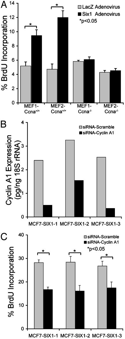
Homeobox genes constitute a large family of transcription factors that are essential during normal development and are often dysregulated in cancer. However, the molecular mechanisms by which homeobox genes influence cancer remain largely unknown. Here we show that the tissue-restricted cyclin A1 is a transcriptional target of the Six1 homeoprotein. Both genes are expressed in the embryonic but not the terminally differentiated mammary gland, and Six1-knockout mice show a dramatic reduction of cyclin A1 in the embryonic mammary gland. In addition, both genes are reexpressed in breast cancers. Six1 overexpression increases cyclin A1 mRNA levels and activity, cell proliferation, and tumor volume, whereas Six1 down-regulation decreases cyclin A1 mRNA levels and proliferation. Overexpression of Six1 in wild-type mouse embryonic fibroblasts, but not in knockout variants lacking the cyclin A1 gene, induces cell proliferation. Furthermore, inhibition of cyclin A1 in Six1-overexpressing mammary carcinoma cells decreases proliferation. Together these results demonstrate that cyclin A1 is required for the proliferative effect of Six1. We conclude that Six1 overexpression reinstates an embryonic pathway of proliferation in breast cancer by up-regulating cyclin A1.


Figures

Fig. 1.
Fig. 1.
Six1 directly activates the tissue-specific cyclin A1. (A) MCF7 cells were transfected with increasing concentrations of the human Six1 expression plasmid (SIXFL) plus the full-length human cyclin A1 promoter–luciferase construct. The relative fold increase in activity is compared with cells transfected with the empty vector plus the cyclin A1 promoter–luciferase construct and with cells transfected with SIXFL and the promotorless luciferase construct, all normalized to Renilla-luciferase activity. (B) Six1 activation of the cyclin A1 promoter in MCF7 cells occurs through the region from -112 to -37. Analysis was performed as described in A, with 5 μg of SIXFL and the promoter–luciferase constructs as depicted in the figure. (C) ChIP assays were performed in 21PT cells with the anti-Six1 antibody (23) or no antibody (control), and the immunoprecipitates were analyzed by PCR with cyclin A1 promoter-specific primers in the Six1 binding region (-207 to -18) or with primers upstream of the binding region (-2312 to -2107).
Fig. 2.
Fig. 2.
Six1 and its target cyclin A1 are reexpressed in human breast cancers. (A) qRT-PCR demonstrates that Six1 and cyclin A1 are highly expressed in the embryonic mammary gland as opposed to the fully differentiated adult pregnant and lactating mammary gland. (B) Reduction of cyclin A1 in Six1-knockout mammary glands. qRT-PCR using probes for Six1 and cyclin A1 was performed on mammary glands isolated from day 18.5 wild-type, heterozygote, and Six1-knockout embryos. Three independent samples for each condition were tested (wild type, heterozygote, and knockout), and each sample represents a pool of at least 15 animals. (C) qRT-PCR demonstrates that Six1 and cyclin A1 mRNA levels are overexpressed in numerous breast cancer cell lines as compared with immortalized normal breast lines. (D) Cyclin A1 levels correlate with Six1 in breast cancer samples as determined by qRT-PCR.
Fig. 3.
Fig. 3.
Overexpression of Six1 induces cyclin A1 expression and cellular proliferation. (A) MCF7-SIX1 transfectants have increased levels of cyclin A1 mRNA as compared with control transfectants. Upper shows a representative immunoprecipitation of Six1 in MCF7-SIX1 and MCF7-Control transfectants, whereas Lower demonstrates by qRT-PCR that cyclin A1 levels are increased in the Six1-transfectants, whereas cyclin A2 levels remain unchanged. (B) Immune-complex kinase assays demonstrate an increase in cyclin A1- and cdk2-associated kinase activities in Six1-overexpressing MCF7 cells, whereas cyclin A2-associated kinase activity remains unchanged. Histone H1 was used as a substrate for all immune complexes. (C and D) Assays measuring cell growth (C) and BrdUrd incorporation (D) demonstrate that Six1 overexpressing cell lines have a statistically significant increase in proliferation as compared with control cells. Each time point in the cell growth curve represents the mean of nine counts for each cell strain and is expressed as the mean ± SD of three cell lines per group. (E) Representative flow cytometry of one Six1-overexpressing (MCF7-SIX1–3) and one control (MCF7-Control 3) transfectant demonstrates that overexpression of Six1 accelerates cell cycle progression in MCF7 cells (similar results were obtained for the other Six1-overexpressing clones). The numbers on the bottom represent hours after release from serum starvation. (F) Expression of Six1 significantly increases tumor burden in nude mice. MCF7 cells (control and Six1-overexpressing) were injected into the flank of 8-week-old nude mice (previously implanted with estrogen pellets, four or five mice per group) at 10 × 106 cells per mouse in 100 μl of medium without serum, and tumor size was measured over a 6-week time period. Data are shown as mean ± SEM.
Fig. 4.
Fig. 4.
Inhibition of Six1 by siRNA in 21PT cells decreases cyclin A1 expression and cellular proliferation. (A) siRNA against Six1 decreases Six1 levels over a 7-day time course. Northern blot (Six1 and actin) and Western blot (Six1) analysis demonstrate that Six1 levels are decreased after introduction of the siRNA-SIX1 construct. For the Western blot analysis, similar protein concentrations were loaded per pair on each day, with increasing concentrations loaded on subsequent days. (B) qRT-PCR and Northern blot analyses demonstrate a decrease in cyclin A1 expression (Upper) when Six1 is inhibited (Lower). (C and D) Cell growth (C) and BrdUrd incorporation (D) assays show a statistically significant decrease in proliferation when Six1 is downregulated by siRNA. (E) Flow cytometry on propidium iodide-stained cells demonstrates a slower progression through the cell cycle when Six1 is downregulated. Inset represents Six1 protein levels over the time course. Numbers represent hours after release from serum starvation.
Fig. 5.
Fig. 5.
Six1 is dependent on cyclin A1 to stimulate cellular proliferation. (A) BrdUrd incorporation assays reveal the dependency of Six1 on cyclin A1 for inducing cellular proliferation, as wild-type cells (MEF-Ccna1+/+) can be stimulated to increase proliferation when Six1 is introduced by adenoviral transduction, whereas cyclin A-deficient cells (MEF-Ccna1-/-) cannot be induced to proliferate by the introduction of Six1. (B) Cyclin A1 mRNA is reduced in Six1-overexpressing MCF7 cells after introduction of a cyclin A1 siRNA to levels at or slightly below levels observed in CAT transfected controls (see Fig. 3A). Cyclin A1 siRNA or a scrambled vector control were introduced into MCF7-SIX1 cells by electroporation. Approximately 36 h after introduction of the siRNA vectors, RNA was isolated from each condition and expression of cyclin A1 was measured by qRT-PCR. (C) Proliferation is decreased in MCF-SIX1 cells when cyclin A1 levels are reduced by means of siRNA technology. Cells were treated as described in B, after which proliferation was measured by BrdUrd incorporation. The experiment was performed twice in triplicate.

References

    1. Abate-Shen, C. (2002) Nat. Rev. Cancer 2, 777-785. - PubMed
    1. Ford, H. L. (1998) Cell Biol. Int. 22, 397-400. - PubMed
    1. Li, X., Perissi, V., Liu, F., Rose, D. W. & Rosenfeld, M. G. (2002) Science 297, 1180-1183. - PubMed
    1. Kawakami, K., Sato, S., Ozaki, H. & Ikeda, K. (2000) BioEssays 22, 616-626. - PubMed
    1. Relaix, F. & Buckingham, M. (1999) Genes Dev. 13, 3171-3178. - PubMed

Publication types

MeSH terms