. 2004 May;113(9):1266-70.
doi: 10.1172/JCI21301.

Hematopoietic myelomonocytic cells are the major source of hepatocyte fusion partners

Affiliations

Hematopoietic myelomonocytic cells are the major source of hepatocyte fusion partners

Fernando D Camargo et al. J Clin Invest. 2004 May.

Abstract

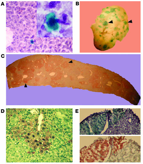
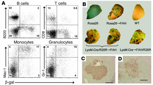
Several recent reports have demonstrated that transplantation of bone marrow cells can result in the generation of functional hepatocytes. Cellular fusion between bone marrow-derived cells and host hepatocytes has been shown to be the mechanism of this phenomenon. However, the exact identity of the bone marrow cells that mediate cellular fusion has remained undetermined. Here we demonstrate that the hematopoietic progeny of a single hematopoietic stem cell (HSC) is sufficient to produce functional hepatic repopulation. Furthermore, transplantation of lymphocyte-deficient bone marrow cells and in vivo fate mapping of the myeloid lineage revealed that HSC-derived hepatocytes are primarily derived from mature myelomonocytic cells. In addition, using a Cre/lox-based strategy, we directly demonstrate that myeloid cells spontaneously fuse with host hepatocytes. Our findings raise the possibility that differentiated myeloid cells may be useful for future therapeutic applications of in vivo cellular fusion.


Figures

Figure 1
Figure 1
Hepatic differentiation after transplantation of a single HSC. Fourteen months after transplantation of a single β-gal+ HSC, random liver sections were analyzed by X-gal staining. (A) Cells with hepatocyte morphology (large cells with abundant cytoplasm) that clearly expressed β-gal and were integrated into the liver parenchyma were detected in primary single-cell recipients. The micrographs were taken at ∞20 magnification, with the inset at ∞100. Clonally derived hematopoietic BM cells were harvested from primary recipients and transplanted into Fah_/_ mice. (B) Five months after transplantation, whole-mount X-gal staining on liver lobes revealed extensive blue nodular repopulation (arrowheads). (C) FAH immunohistochemistry (dark brown staining) shown on a liver section confirms widespread oligoclonal repopulation throughout the host parenchyma (arrowheads). (D) Increased magnification of a repopulated nodule reveals typical hepatocyte morphology of the brown FAH+ cells. (E) Frozen serial sections stained with X-gal and anti-FAH show colocalization of staining, confirming that the FAH expression is derived from the original Rosa26 (lacZ+) HSC.
Figure 2
Figure 2
Myeloid-derived hepatocyte regeneration in Fah_/_ hosts. (A) Cytofluorimetric analysis of peripheral blood from FAH mutant mice transplanted with LysM-Cre/R26R BM. The flow profiles shown were previously gated on CD45.2+ cells in order to restrict analysis to donor-derived cells. B220 was used as a B cell antibody, CD8 as a T cell antibody, and Gr-1 and Mac-1 as myeloid cell antibodies. (B) Histochemical detection of β-gal activity in Fah_/_ recipient livers and controls. Rosa26 and wild-type livers were collected from untransplanted mice of the respective genotype. In the case of transplanted mice, the genotype preceding the arrow represents the donor WBM genotype. Host genotype is indicated after the arrow. (C and D) FAH immunohistochemistry on liver sections of recipients of LysM-Cre/R26R (C) and LysM-Cre (D) WBM, indicating donor-derived hepatocyte nodules. Scale bars: 200 ∝m.

Comment in

  • J Clin Invest. 113:1249.

References

    1. Ferrari G, et al. Muscle regeneration by bone marrow-derived myogenic progenitors. Science. 1998;279:1528–1530. - PubMed
    1. Jackson KA, et al. Regeneration of ischemic cardiac muscle and vascular endothelium by adult stem cells. J. Clin. Invest. 2001;107:1395–1402. - PMC - PubMed
    1. Gussoni E, et al. Dystrophin expression in the mdx mouse restored by stem cell transplantation. Nature. 1999;401:390–394. - PubMed
    1. Asahara T, et al. Bone marrow origin of endothelial progenitor cells responsible for postnatal vasculogenesis in physiological and pathological neovascularization. Circ. Res. 1999;85:221–228. - PubMed
    1. Mezey E, Chandross KJ, Harta G, Maki RA, McKercher SR. Turning blood into brain: cells bearing neuronal antigens generated in vivo from bone marrow. Science. 2000;290:1779–1782. - PubMed

Publication types

Substances