. 2004 May;113(9):1296-306.
doi: 10.1172/JCI20295.

Bacterial flagellin is a dominant antigen in Crohn disease

Affiliations

Bacterial flagellin is a dominant antigen in Crohn disease

Michael J Lodes et al. J Clin Invest. 2004 May.

Abstract

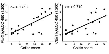
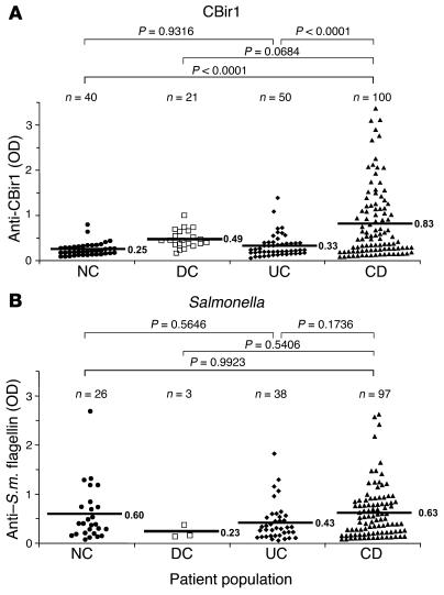
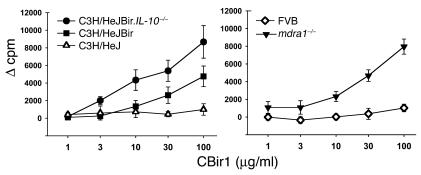
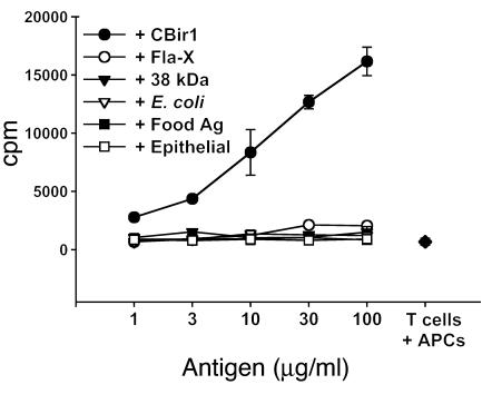
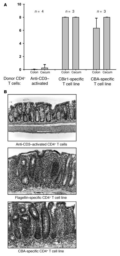
Chronic intestinal inflammation, as seen in inflammatory bowel disease (IBD), results from an aberrant and poorly understood mucosal immune response to the microbiota of the gastrointestinal tract in genetically susceptible individuals. Here we used serological expression cloning to identify commensal bacterial proteins that could contribute to the pathogenesis of IBD. The dominant antigens identified were flagellins, molecules known to activate innate immunity via Toll-like receptor 5 (TLR5), and critical targets of the acquired immune system in host defense. Multiple strains of colitic mice had elevated serum anti-flagellin IgG2a responses and Th1 T cell responses to flagellin. In addition, flagellin-specific CD4(+) T cells induced severe colitis when adoptively transferred into naive SCID mice. Serum IgG to these flagellins, but not to the dissimilar Salmonella muenchen flagellin, was elevated in patients with Crohn disease, but not in patients with ulcerative colitis or in controls. These results identify flagellins as a class of immunodominant antigens that stimulate pathogenic intestinal immune reactions in genetically diverse hosts and suggest new avenues for the diagnosis and antigen-directed therapy of patients with IBD.


Figures

Figure 1
Figure 1
Flagellin clone identity and similarity to known flagellin sequences. (A) Schematic of CBir flagellin clones from serological expression screening. The predicted amino acid sequences from the flagellin expression clones (CBir1_CBir15) are mapped in relation to the representation of the B. fibrisolvens sequence at the top. Ruler length equals 500 amino acids. Similarity in the NH2 conserved sequence between these flagellin clones and B. fibrisolvens sequences ranged from 45 to 84% (mean, 60.3%). Breaks in the lines representing clones CBir1 and CBir2 indicate differences in sequence length in the hypervariable region. NH2-conserved, conserved NH2 sequence; CO2H-conserved, conserved carboxy sequence. (B) Phylogenetic tree showing relatedness at the conserved NH2 termini of CBir1_CBir15 clones to flagellin sequences in the GenBank database. The dendrogram was constructed using the Clustal program in DNAStar and reflects similarity at the amino acid level. The approximate location of the Clostridium subphylum cluster XIVa is indicated with a bracket.
Figure 2
Figure 2
Schematic of recombinant flagellin constructs with percent similarity to related flagellin B from the anaerobe B. fibrisolvens (GenBank accession number AAB82613). (A) Structure of B. fibrisolvens flagellin B showing conserved NH2 and carboxy (CO2H-conserved) regions and the hypervariable central domain. (B) Diagram of the full-length amino acid sequence of mouse cecal bacteria flagellins CBir1 and Fla-X, indicating the similarity of the three domains with the respective B. fibrisolvens domains. (C and D) Schematics of recombinant flagellin proteins and fragments for CBir1 (C) and Fla-X (D) expressed in E. coli and purified by six-histidine tag affinity to nickel-nitrilotriacetic acid columns.
Figure 3
Figure 3
Western blot analysis of the serum antibody response to recombinant flagellins CBir1 and Fla-X and their fragments. (A) Noncolitic C3H/HeJ (pool of two) versus colitic C3H/HeJBir (pool of five) mice. (B) Noncolitic FVB (pool of five) versus colitic mdr1a–/– (pool of five) mice. (C) Random human blood donor (Normal human) versus a pool of CD patients with severe disease. Protein samples include mouse CBA, full-length recombinant proteins (FL), the NH2 conserved region (A) and the conserved carboxy region (C) of flagellin (see Figure 2, C and D).
Figure 4
Figure 4
ELISA titration of mouse serum anti-flagellin against recombinant flagellins CBir1 and Fla-X with secondary antibodies specific for mouse IgG, IgG1, and IgG2a antibodies. Colitic C3H/HeJBir serum (pool of five) versus noncolitic C3H/HeJ serum (pool of two) was used in the upper panel and colitic mdr1a–/– serum (pool of five) versus noncolitic FVB serum (pool of five) was used in the lower panel.
Figure 5
Figure 5
Correlation of colitis histopathology score (0_60) with serum anti_Fla-X and anti-CBir1. Twenty-three mdr1a–/– mice, ranging in age from 6 to 13 weeks, were randomly chosen for assignment of quantitative histopathology scores. Serum anti-flagellin from these mice was quantified by ELISA. Colitis scores of 0_2 represent no disease; 3_15, mild disease, 16_35, moderate disease, and more than 35, severe disease (22). Similar results were obtained for both recombinant flagellins: Fla-X (left panel) and CBir1 (right panel).
Figure 6
Figure 6
Association of anti-flagellin antibodies with human IBDs. Human sera, well characterized for CD and UC, were tested by ELISA for reactivity to flagellin CBir1 (A) and Salmonella muenchen (S.m.) flagellin (B). Statistical analysis was performed with the Tukey-Kramer test; the resulting statistics (P values) as well as population size (n) are shown above the graphs. Mean OD450 values are indicated by horizontal bars.
Figure 7
Figure 7
Dose response of CD4+ T cell proliferation to CBir1 and Fla-X in multiple strains of mice. Left panel: C3H/HeJ (open triangles), C3H/HeJBir (squares), and C3H/HeJBir.IL-10–/– (circles). Right panel: FVB (diamonds) and mdr1a–/– (filled triangles). The y axes indicate sample counts per minute (cpm) minus control T cell plus APC cpm (Ø cpm) for each experimental group. The x axes indicate the dose (∝g/ml) of recombinant flagellin used in each assay. Vertical bars indicate plus or minus one standard deviation of the mean value.
Figure 8
Figure 8
Dose response and specificity of C3H/HeJBir CD4+ CBir1-specific T cell line. T cell line CBir-1B1 proliferated specifically in response to recombinant flagellin protein CBir1. Antigens used in the assay include recombinant flagellins CBir1 (filled circles) and Fla-X (open circles); the 38-kDa antigen of M. tuberculosis (p38 antigen; 38 kDa: filled triangles); lysate of E. coli antigens (E. coli; open triangles); protein antigens extracted from mouse food pellets (Food Ag; filled squares); and a lysate of the ModeK epithelial cell line, of C3H origin (epithelial: open squares). Several randomly expressed recombinant commensal bacterial antigens were also tested and were negative (including randomly cloned C3H/HeJ mouse cecal bacterial antigens 99 [rIB99] and 32 [rIB32]). T cells plus APCs only are indicated by a filled diamond.
Figure 9
Figure 9
Adoptive transfer of C3H/HeJBir CD4+ CBir1-specific T cell line into C3H/HeJ scid/scid recipients. (A) Two months after transfer, cecal and colon histopathology was assigned scores with a quantitative system (14). CD4+ T cells activated polyclonally with mAb against CD3 prior to transfer were used as a negative control (Anti-CD3_activated). A CBA-specific CD4+ T cell line reactive with unselected cecal bacterial antigens was used as a positive control (CBA-specific T cell line); the CBir1-specific CD4+ T cell line corresponds to the flagellin-specific T cell line in Figure 8. Sample size (n) is indicated at the top. (B) Representative histopathology of the groups shown in A: Anti-CD3_activated CD4+ T cells (top panel), CBir1 flagellin_specific CD4+ T cells (middle panel), and CBA-specific CD4+ T cells (bottom panel). Magnification, ∞200.

References

    1. Strober W, Fuss IJ, Blumberg RS. The immunology of mucosal models of inflammation. Annu. Rev. Immunol. 2002;20:495–549. - PubMed
    1. Bouma G, Strober W. The immunological and genetic basis of inflammatory bowel disease. Nat. Rev. Immunol. 2003;3:521–533. - PubMed
    1. McCracken VJ, Lorenz RG. The gastrointestinal ecosystem: a precarious alliance among epithelium, immunity and microbiota. Cell. Microbiol. 2001;3:1–11. - PubMed
    1. Kopp E, Medzhitov R. Recognition of microbial infection by Toll-like receptors. Curr. Opin. Immunol. 2003;15:396–401. - PubMed
    1. Akira S. Mammalian Toll-like receptors. Curr. Opin. Immunol. 2003;15:5–11. - PubMed

Publication types

MeSH terms

Associated data