Comparative Study
. 2004 May;113(9):1318-27.
doi: 10.1172/JCI19930.

Pre-B cell colony-enhancing factor inhibits neutrophil apoptosis in experimental inflammation and clinical sepsis

Affiliations
Comparative Study

Pre-B cell colony-enhancing factor inhibits neutrophil apoptosis in experimental inflammation and clinical sepsis

Song Hui Jia et al. J Clin Invest. 2004 May.

Abstract

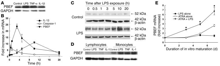
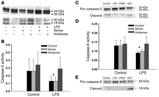
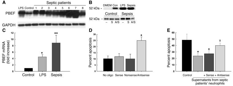
Pre-B cell colony-enhancing factor (PBEF) is a highly conserved 52-kDa protein, originally identified as a growth factor for early stage B cells. We show here that PBEF is also upregulated in neutrophils by IL-1beta and functions as a novel inhibitor of apoptosis in response to a variety of inflammatory stimuli. Induction of PBEF occurs 5-10 hours after LPS exposure. Prevention of PBEF translation with an antisense oligonucleotide completely abrogates the inhibitory effects of LPS, IL-1, GM-CSF, IL-8, and TNF-alpha on neutrophil apoptosis. Immunoreactive PBEF is detectable in culture supernatants from LPS-stimulated neutrophils, and a recombinant PBEF fusion protein inhibits neutrophil apoptosis. PBEF is also expressed in neutrophils from critically ill patients with sepsis in whom rates of apoptosis are profoundly delayed. Expression occurs at higher levels than those seen in experimental inflammation, and a PBEF antisense oligonucleotide significantly restores the normal kinetics of apoptosis in septic polymorphonuclear neutrophils. Inhibition of apoptosis by PBEF is associated with reduced activity of caspases-8 and -3, but not caspase-9. These data identify PBEF as a novel inflammatory cytokine that plays a requisite role in the delayed neutrophil apoptosis of clinical and experimental sepsis.


Figures

Figure 1
Figure 1
PBEF is expressed in neutrophils and monocytes exposed to inflammatory stimuli. (A) Neutrophils expressed mRNA transcripts for PBEF in response to LPS, TNF-α, and IL-1β, stimuli that inhibit neutrophil apoptosis. (B) LPS (1 ∝g/ml) induced transcripts for caspase-1 (open circles), reaching maximal concentrations by 1 hour, IL-1β (filled circles) peaking at 3 hours, and PBEF (filled triangles) reaching maximal levels at 10 hours. The mRNA expression was quantified by real-time PCR; data are expressed as fold increase over basal levels of expression for each mRNA species; n = 4. (C) Western blot analysis showed low-level expression of PBEF in control cells, while LPS (1 ∝g/ml) increased PBEF protein, with maximum expression evident 10 hours after exposure. Blots were reprobed with β-actin to confirm equal loading; data are representative of three separate experiments. (D) PBEF mRNA transcripts were also expressed in peripheral blood monocytes, but not lymphocytes, in response to LPS, TNF, and IL-1. Blots are representative of three separate experiments; corresponding mRNA for GAPDH is shown to evaluate sample loading. (E) HL-60 cells induced to granulocytic differentiation by 1 ∝M all-trans retinoic acid showed increased message for PBEF, and transcript levels evaluated by real-time PCR were further increased by exposure of differentiated HL-60 cells to LPS (1 ∝g/ml) added to cultures with all-trans retinoic acid at day 0. Data are normalized to levels of PBEF transcripts in undifferentiated HL-60 cells; *P < 0.05 versus all-trans retinoic acid alone; n = 4. ATRA, all-trans retinoic acid.
Figure 2
Figure 2
Prevention of PBEF translation with an antisense oligonucleotide blocks inhibition of apoptosis in response to inflammatory stimuli. (A) Neutrophil apoptosis, assessed as the nuclear uptake of propidium iodide, was significantly inhibited by coincubation with LPS (1 ∝g/ml), IL-1β (100 pg/ml), GM-CSF (20 ng/ml), IL-8 (250 ng/ml), or TNF-α (40 ng/ml). *P < 0.05 compared with control rates. Prevention of PBEF translation using an antisense oligonucleotide prevented this inhibitory effect; the corresponding sense or scrambled nonsense controls were without effect. Data are mean ± SD of six separate studies. (B) Antisense treatment of resting or LPS-stimulated (1 ∝g/ml) neutrophils inhibited the translation of PBEF as detected by Western blot analysis. Blot is representative of three separate studies. (C) Both LPS (1 ∝g/ml) and recombinant PBEF (50 ng/ml) inhibited phosphatidylserine exteriorization detected by the binding of FITC-labeled annexin V, an effect that was specifically blocked when neutrophils were pretreated with PBEF antisense; n = 4, *P < 0.05. rPBEF, recombinant PBEF.
Figure 3
Figure 3
PBEF exerts its antiapoptotic activity as a secreted factor. (A) Naive neutrophils were incubated for 21 hours with conditioned medium from resting or LPS-stimulated neutrophils, with or without prior transfection with PBEF antisense oligonucleotide or the sense control. Conditioned medium from resting neutrophils did not alter apoptotic rates in comparison with untreated controls (white bar). Conditioned medium from neutrophils incubated with LPS inhibited the apoptosis of naive neutrophils (*P < 0.05 versus control cell supernatants). Pretreatment with an antisense PBEF oligonucleotide, but not with the sense control, blocked this inhibitory activity (#P < 0.05 versus sense control). Data are means ± SD of n = 6 experiments. (B) Culture supernatants or whole cell lysates from CHO cells transfected with a pCDNA3.1 vector carrying a PBEF/c-myc construct were immunoprecipitated with anti_c-myc. PBEF could be detected by Western blot analysis with anti-PBEF Ab in both lysates and supernatants from transfected cells, but not in those from nontransfected cells or cells transfected with plasmid containing c-myc alone. (C) Supernatants from transfected CHO cells suppressed the apoptosis of resting neutrophils as measured by propidium iodide uptake, whereas supernatants from vector-treated controls were without effect; *P < 0.05 versus control or empty vector, n = 4. (D) A recombinant PBEF/GST fusion protein added to cultures of control neutrophils induced dose-dependent inhibition of apoptosis; polymyxin B (10 ∝g/ml) was added to cultures to neutralize any contaminating LPS. Recombinant GST alone was without effect (data not shown). Apoptosis was measured by propidium iodide uptake at 21 hours; results are means ± SD of n = 3 studies.
Figure 4
Figure 4
Both extracellular and endogenous PBEF contribute to the inhibition of neutrophil apoptosis. (A) Polyclonal anti-PBEF Ab (1:500) added to the culture medium neutralized the antiapoptotic effects of recombinant PBEF/GST, but in contrast to the effects of inhibition of PBEF translation with antisense, oligonucleotide did not block the antiapoptotic effects of LPS; n = 4, *P < 0.05 versus controls. (B) Anti-PBEF Ab (1:500), however, significantly blocked the inhibitory activity of conditioned medium (CM) from LPS-treated neutrophils. Mean ± SD, n = 3, *P < 0.05 versus controls, **P < 0.05 versus conditioned medium alone (no anti-PBEF Ab added). (C) While antisense pretreatment prevented the inhibitory effects of LPS exposure, addition of inhibitory concentrations of rPBEF/GST was unable to restore the inhibitory effect, suggesting that inhibition of apoptosis requires both extracellular and intracellular or endogenous PBEF; n = 3.
Figure 5
Figure 5
PBEF inhibits the cleavage and catalytic activity of caspase-3 and blocks the activity of caspase-8. (A) Caspase-3 is cleaved to yield active 18- to 20-kDa fragments in constitutively apoptotic neutrophils; LPS inhibits the activational cleavage of this effector caspase, as evaluated by Western blot analysis following 6 hours of in vitro culture. PBEF antisense prevented the inhibitory effects of LPS, while the sense control did not. Blot shown is representative of three separate studies. (B) Caspase-3 activity, measured colorimetrically in arbitrary units as the cleavage of the tetrapeptide Ac-DEVD-pNA was also reduced in neutrophils following exposure to LPS; PBEF antisense, but not the sense control, prevented this reduction in caspase-3 activity. Results are means ± SD of six separate studies; *P < 0.05 versus control levels or activity levels in neutrophils treated with PBEF antisense. (C) Cleavage of pro_caspase-3 to active caspase-3 (20 kDa) was reduced in neutrophils incubated with LPS (1 μg/ml) or rPBEF (50 ng/ml); Western blot is representative of 3 separate experiments. (D) Caspase-8 activity, measured colorimetrically in arbitrary units as the cleavage of the caspase-8 tetrapeptide target Ac-IETD-pNA was reduced by exposure to LPS and restored by pretreatment of neutrophils with PBEF antisense. Results are means ± SD of six separate studies; *P < 0.05, control or sense-treated cells versus antisense-treated neutrophils or neutrophils cultured in the absence of LPS. (E) Cleavage of pro_caspase-8 (53 kDa) to its active form (18 kDa) was also inhibited by exposure of neutrophils to LPS or rPBEF; Western blot is representative of 3 separate experiments.
Figure 6
Figure 6
PBEF is expressed and is biologically active in neutrophils harvested from critically ill septic patients. (A) PBEF mRNA in neutrophils from eight critically ill septic patients was expressed at higher levels than in control (Con) or LPS-stimulated neutrophils. Blots were reprobed with GAPDH to confirm comparability of loading. (B) Expression of PBEF mRNA transcripts in septic and LPS-treated neutrophils was evaluated by real-time PCR, normalizing expression to that for GAPDH. Expression was induced by LPS (*P < 0.05 versus unstimulated cells) and even more in septic neutrophils (**P < 0.05 versus both LPS-stimulated cells and unstimulated cells). (C) Immunoreactive PBEF was detectable by Western blot in supernatants from LPS-treated and septic neutrophils following 21 hours of in vitro culture in serum-free medium; antisense pretreatment blocked the secretion of PBEF. DMEM denotes medium only; studies were repeated three times, and a representative blot is shown. S, sense; A/S, antisense. (D) Neutrophils from 16 septic critically ill patients were incubated for 5 hours with PBEF antisense or the sense or nonsense controls, and apoptosis was evaluated 21 hours later. Antisense treated cells, but not controls, showed increased rates of apoptosis (*P = 0.002 versus no oligonucleotide [no oligo]; ANOVA). (E) Supernatants from control PMN had minimal effects on the apoptosis of resting PMN (black bar). In contrast, supernatants from septic PMN or septic PMN incubated with PBEF sense oligonucleotides, significantly inhibited the apoptosis of control PMN (*P < 0.05), whereas supernatants from antisense-treated septic PMNs induced significantly less inhibition ( P < 0.05 versus sense or no oligonucleotide; P = NS versus controls, n = 5).

References

    1. Riedemann NC, Guo R-F, Ward PA. The enigma of sepsis. J. Clin. Invest. 2003;112:460–467. doi:10.1172/JCI200319523. - PMC - PubMed
    1. Cohen J. The immunopathogenesis of sepsis. Nature. 2002;420:885–891. - PubMed
    1. Angus DC, et al. Epidemiology of severe sepsis in the United States: analysis of incidence, outcome, and associated costs of care. Crit. Care Med. 2001;29:1303–1310. - PubMed
    1. Bone RC, et al. Definitions for sepsis and organ failure and guidelines for the use of innovative therapies in sepsis. Chest. 1992;101:1644–1655. - PubMed
    1. Lekstrom-Himes JA, Gallin JI. Immunodeficiency diseases caused by defects in phagocytes. N. Engl. J. Med. 2000;343:1703–1714. - PubMed

Publication types

MeSH terms