

Salivary gland tumors in Uganda: clinical pathological study
- PMID: 15126188
- PMCID: PMC2141656
Salivary gland tumors in Uganda: clinical pathological study
Abstract
Background: The incidence of salivary gland tumors is claimed to be influenced by geographical and racial factors. The pathological classification and nomenclature of salivary gland tumors as defined by WHO classification (1991), is accepted world-wide but little is available in the literature regarding the spectrum of salivary gland tumors in Africa in the basis of this classification. Such efforts would allow comparison and justify any differences between the black African population and the rest of the world.
Objective: To outline the clinicopathological features of salivary gland tumors in Uganda.
Setting: Makerere University, Faculty of Medicine, Department of Pathology.
Methods: All epithelial tumors from major and minor salivary glands accessioned from 1979 to 1988 were analyzed in respect to sex and age of patients, anatomical location of the tumor and histological type. The histological diagnosis of each individual tumor was based on the 1991 WHO classification of salivary gland tumors.
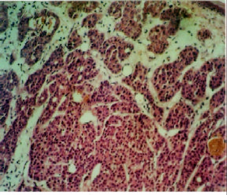
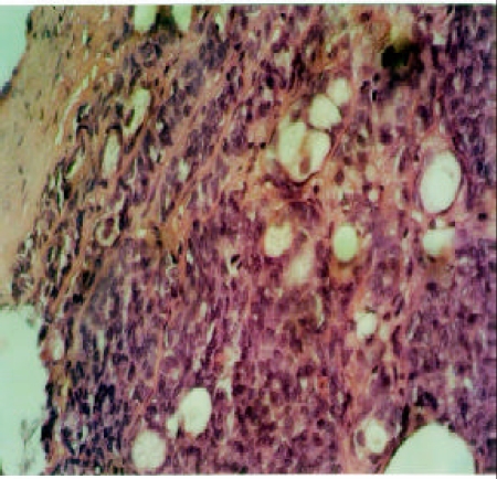
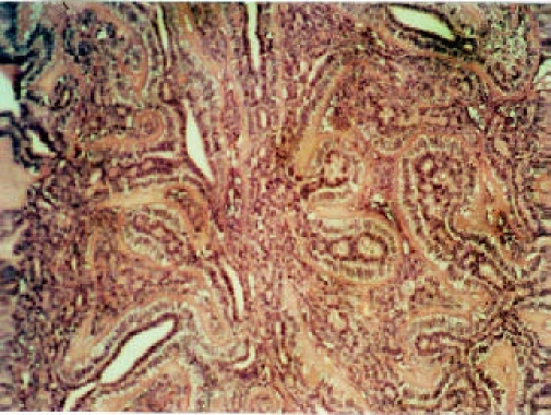
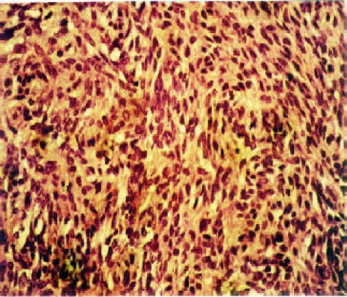
Results: During the span of 10 years, 268 cases of salivary gland tumors were diagnosed. Of these, 113 (42.2%) were males, 148 (55.2%) females and in the remaining seven (2.6%) cases, the sex was not specified. The age range of the 247 patients with recorded ages was from 0.5 to 80 years. The mean age at diagnosis was 38.1 (SD =17.03) with the median of 38.0 years. Thirty four percent of tumors originated from the parotid, 33.2% from the submandibular and 32.8% from minor salivary glands. No tumor was implicated from the sublingual gland. There were a total of 125 (46.6%) malignant tumors and 143 (53.4%) benign tumors. The mean age of patients with malignant lesions (43.1 years; SD=16.75; median=44.00 years) was 9.6 years older than those with benign tumors (mean=33.5 years; SD=16.0; median=30.00 years). Pleomorphic adenoma was the most common benign tumor (74.8%), followed by myoepithelioma (9.8%). No Whartin's tumor was encountered. The malignant tumors were dominated by adenoid cystic carcinoma (28.8%) followed by mucoepidermoid carcinoma (21.6%).
Conclusion: The pattern of distribution of salivary gland tumors in black African population seems to differ from that of Western series in that; i) females are more affected than males, ii) there is a low proportion of tumors from the parotid gland and high proportion of tumors from the submandibular and minor salivary glands, iii) the parotid and minor salivary gland tumors have more probability of being malignant than those tumors from the submandibular gland iv) the newly categorized pathological entities are common and v) Whartin's tumor is extremely rare in black African population.
Figures
References
-
- Seifert G, Miehlke A, Haubrich J, Chilla R. Disease of the Salivary Glands: Pathology-Diagnosis-treatment-Facial Nerve Surg. New York: Georg Thieme Verlag Inc. Stuttgart; 1986.
-
- Eveson JW, Cawson RA. Salivary gland tumors. A review of 2410 cases with particular reference to histological types, site, age and sex distribution. Journal of Pathology. 1985;146:51–58. - PubMed
-
- Sharkey FE. Systematic evaluation of the World Health Organization Classification of salivary gland tumors: clinicopathological study of 366 cases. Amer J Clin Pathol. 1977;67:272–278. - PubMed
-
- Thomas KM, Borgstein J. Salivary gland tumors in Malawi. Cancer. 1980;46:2328–2334. - PubMed
-
- Davies JNP, Dodge HD, Burkitt D. Salivary tumors in Uganda. Cancer. 1964;17:1310–1322. - PubMed
Publication types
MeSH terms
LinkOut - more resources
Full Text Sources
Medical