. 2004 May;186(10):3202-13.
doi: 10.1128/JB.186.10.3202-3213.2004.

Precise excision of the large pathogenicity island, SPI7, in Salmonella enterica serovar Typhi

Affiliations

Precise excision of the large pathogenicity island, SPI7, in Salmonella enterica serovar Typhi

Susan M Bueno et al. J Bacteriol. 2004 May.

Abstract

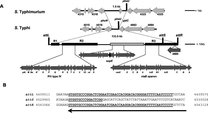
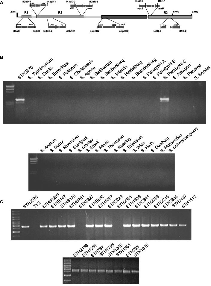
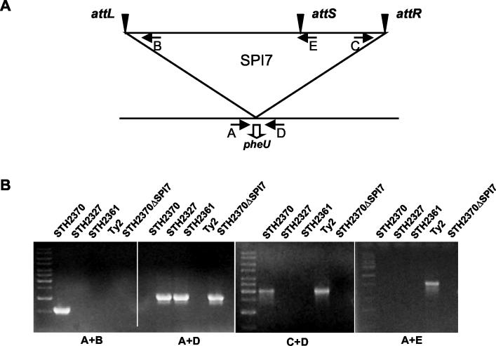
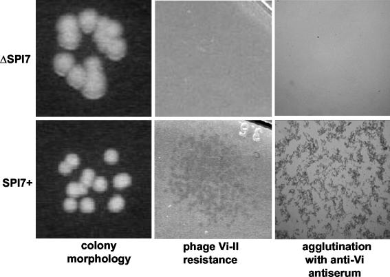
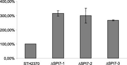
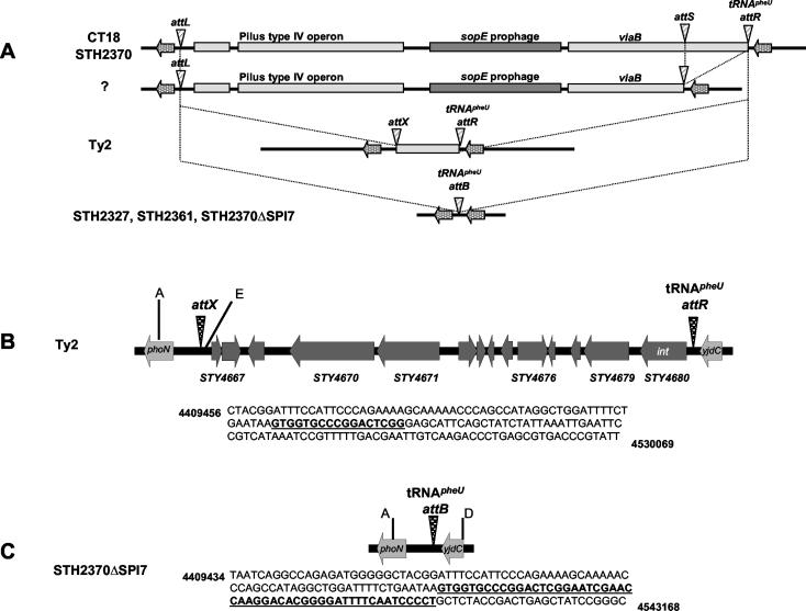
The large pathogenicity island (SPI7) of Salmonella enterica serovar Typhi is a 133,477-bp segment of DNA flanked by two 52-bp direct repeats overlapping the pheU (phenylalanyl-tRNA) gene, contains 151 potential open reading frames, and includes the viaB operon involved in the synthesis of Vi antigen. Some clinical isolates of S. enterica serovar Typhi are missing the entire SPI7, due to its precise excision; these strains have lost the ability to produce Vi antigen, are resistant to phage Vi-II, and invade a human epithelial cell line more rapidly. Excision of SPI7 occurs spontaneously in a clinical isolate of S. enterica serovar Typhi when it is grown in the laboratory, leaves an intact copy of the pheU gene at its novel join point, and results in the same three phenotypic consequences. SPI7 is an unstable genetic element, probably an intermediate in the pathway of lateral transfer of such pathogenicity islands among enteric gram-negative bacteria.


Figures

FIG. 1.
FIG. 1.
Structure of S. enterica serovar Typhi SPI7. (A) Alignment of the segment of the S. enterica serovar Typhi CT18 genome sequence (45) containing SPI7 with the corresponding segment of the S. enterica serovar Typhimurium LT2 genome (37). Gene numbers (STM and STY, respectively) are as designated in the annotations of these genome sequences. In place of the 133.5-kb SPI7 in the S. enterica serovar Typhi genome, S. enterica serovar Typhimurium has a 1.5-kb segment of DNA. SPI7 is flanked on the left by the phoN gene and the incomplete 3′ end of the pheU gene and on the right by the complete pheU and adjacent yjdC genes; other gene numbers shown are from the annotated genome sequences. SPI7 has three regions of genes, R1, R2, and R3 (solid boxes), that are not found in other serovars of S. enterica, with the exception of serovar Paratyphi C (see the text). Region 2 includes the genes involved in the synthesis of type IV pili (pilL-pilK), and region 3 includes the Vi antigen biosynthetic (viaB) operon (vexE-tviA). A copy of the sopE gene is found between regions 2 and 3, flanked by genes similar to a subset of phage P2 genes. As described in the text, this region also includes genes predicted to encode homologues of plasmid transfer factors and of phage P4 proteins. Among the genes similar to those of phage P4 is STY4680, located adjacent to the right end (attR) of SPI7 and predicted to encode an integrase. (B) Comparison of the 52-bp left and right repeat sequences (attL and attR) flanking SPI7 with the short 17-bp repeat sequence (attS) found downstream of the viaB operon. The arrow at the bottom indicates the 3′ end of the pheU gene; coordinates (given as base pairs in the text) are those of the S. enterica serovar Typhi CT18 sequence (45).
FIG. 2.
FIG. 2.
SPI7 is found in only two serovars of S. enterica, Typhi and Paratyphi C, and is not present in a subset of S. enterica serovar Typhi clinical isolates. (A) Positions of the primers used to amplify subregions of SPI7. Each primer pair (D [direct] and R [reverse]) was designed to amplify a <3-kb subregion of SPI7 present in the S. enterica serovar Typhi CT18 sequence (45); positive results for the majority of these amplifications show that SPI7 is present in most clinical isolates of S. enterica serovar Typhi used in this study. (B) PCR was used to amplify a region internal to region 2 of SPI7 (bp 4454572 to 4457403) in isolates of different serovars of S. enterica. Only isolates of serovars Typhi and Paratyphi C yielded products after amplification with this primer pair corresponding to the S. enterica serovar Typhi CT18 sequence. (C) SPI7 is found in a majority of clinical isolates of S. enterica serovar Typhi. Similar results were obtained with five additional primer pairs designed to amplify subregions of regions 1, 2, and 3 (data not shown).
FIG. 3.
FIG. 3.
Clinical isolates of S. enterica serovar Typhi missing a region internal to SPI7 have undergone precise deletion of SPI7. The PCR strategy for this experiment is diagrammed at the top and is described in the text. The results are shown at the bottom.
FIG. 4.
FIG. 4.
Precise excision of SPI7 results in profound phenotypic changes. Shown are differences in the colony morphologies (×2 magnification) (left), sensitivities to phage Vi-II (×2 magnification) (center), and agglutination with anti-Vi antiserum (×10 magnification) (right) exhibited by otherwise isogenic derivatives of S. enterica serovar Typhi clinical isolate STH2370 with (SPI7+) and without (ΔSPI7) SPI7.
FIG. 5.
FIG. 5.
Derivatives of STH2370 that have lost SPI7 are hyperinvasive in human HEp-2 cells. The titers of mutant intracellular bacteria after 1 h relative to that of the wild-type strain are a measure of the ability of each mutant to invade epithelial cells; under these conditions, about 0.2 to 0.4% of the infecting STH2370 bacteria can be recovered from human cells after gentamicin treatment. The figure shows the average results ± standard deviations from three independent determinations for three independent derivatives of STH2370 that have lost SPI7 by precise excision, as described in Materials and Methods.
FIG. 6.
FIG. 6.
Novel join points formed by site-specific recombination between the attL, attS, and attR sites of SPI7. (A) Structure of SPI7 found in the majority of clinical isolates of S. enterica serovar Typhi, including CT18 and STH2370. Although we could not detect whether attS × attR recombination could occur in strains of S. enterica serovar Typhi, site-specific recombination between attL and attS has occurred in our working strain of Ty2, to give rise to the novel join point, attX, and between attL and attR in clinical isolates STH2327 and STH2361, to give rise to the novel join point, attB. This is the identical join point to that found in derivatives of STH2370. (B and C) Detailed structures of the chromosomal products of site-specific recombination within SPI7 in strains Ty2 and STH2370ΔSPI7, as explained in the legend to Fig. 1, as well as the sequences of the novel join points attX and attB, respectively. Coordinates are those of the S. enterica serovar Typhi CT18 sequence (45).

References

    1. Arricau, N., D. Hermant, H. Waxin, C. Ecobichon, P. S. Duffey, and M. Y. Popoff. 1998. The RcsB-RcsC regulatory system of Salmonella typhi differentially modulates the expression of invasion proteins, flagellin and Vi antigen in response to osmolarity. Mol. Microbiol. 29:835-850. - PubMed
    1. Bach, S., C. Buchrieser, M. Prentice, A. Guiyoule, T. Msadek, and E. Carniel. 1999. The high-pathogenicity island of Yersinia enterocolitica Ye8081 undergoes low-frequency deletion but not precise excision, suggesting recent stabilization in the genome. Infect. Immun. 67:5091-5099. - PMC - PubMed
    1. Bakshi, C. S., V. P. Singh, M. W. Wood, P. W. Jones, T. S. Wallis, and E. E. Galyov. 2000. Identification of SopE2, a Salmonella secreted protein which is highly homologous to SopE and involved in bacterial invasion of epithelial cells. J. Bacteriol. 182:2341-2344. - PMC - PubMed
    1. Blum, G., M. Ott, A. Lischewski, A. Ritter, H. Imrich, H. Tsachape, and J. Hacker. 1994. Excision of large DNA regions termed pathogenicity island from tRNA-specific loci in the chromosome of an Escherichia coli wild-type pathogen. Infect. Inmmun. 62:606-614. - PMC - PubMed
    1. Bossi, L., J. A. Fuentes, G. Mora, and N. Figueroa-Bossi. 2003. Prophage contribution to bacterial population dynamics. J. Bacteriol. 185:6467-6471. - PMC - PubMed

Publication types

Substances

LinkOut - more resources