Comparative Study
. 2004 Jul 1;381(Pt 1):13-8.
doi: 10.1042/BJ20040434.

TIRF imaging of docking and fusion of single insulin granule motion in primary rat pancreatic beta-cells: different behaviour of granule motion between normal and Goto-Kakizaki diabetic rat beta-cells

Affiliations
Comparative Study

TIRF imaging of docking and fusion of single insulin granule motion in primary rat pancreatic beta-cells: different behaviour of granule motion between normal and Goto-Kakizaki diabetic rat beta-cells

Mica Ohara-Imaizumi et al. Biochem J. .

Abstract

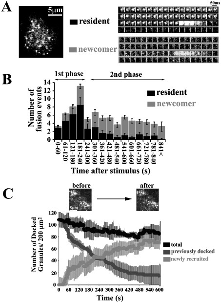
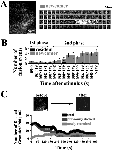
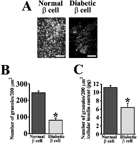
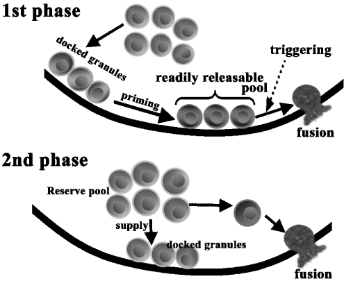
We imaged and analysed the motion of single insulin secretory granules near the plasma membrane in live pancreatic beta-cells, from normal and diabetic Goto-Kakizaki (GK) rats, using total internal reflection fluorescence microscopy (TIRFM). In normal rat primary beta-cells, the granules that were fusing during the first phase originate from previously docked granules, and those during the second phase originate from 'newcomers'. In diabetic GK rat beta-cells, the number of fusion events from previously docked granules were markedly reduced, and, in contrast, the fusion from newcomers was still preserved. The dynamic change in the number of docked insulin granules showed that, in GK rat beta-cells, the total number of docked insulin granules was markedly decreased to 35% of the initial number after glucose stimulation. Immunohistochemistry with anti-insulin antibody observed by TIRFM showed that GK rat beta-cells had a marked decline of endogenous insulin granules docked to the plasma membrane. Thus our results indicate that the decreased number of docked insulin granules accounts for the impaired insulin release during the first phase of insulin release in diabetic GK rat beta-cells.


Figures

Figure 1
Figure 1. TIRF images and analysis of single GFP-labelled insulin granule motion in normal rat primary β-cells during glucose-induced biphasic insulin release
(A) The real-time motion of GFP-labelled insulin granules was imaged close to the plasma membrane (50-ms intervals) (see Supplemental Movie 1, http://www.BiochemJ.org/bj/381/bj3810013add.htm). Sequential images (1 μm×1 μm) of a granule docking and fusing with the plasma membrane were presented during stimulation with 22 mM glucose. ‘Resident’ indicates that the morphologically previously docked granule is fused with the plasma membrane. Fusion is observed as the rapid spread of brightened fluorescence, followed by its disappearance. ‘Newcomer’ indicates that the granules approach from the inside (being absent before stimulation with 22 mM glucose), reach the plasmalemma and then are quickly fused. (B) Histogram showing the number of fusion events (per 200 μm2) at 60-s intervals after stimulation (n=10 cells). The black columns show the fusion from residents, and the grey columns shows that from newcomers. During the first phase, fusion occurs mostly from residents. The fusing granules during the second phase originate mostly from newcomers. (C) Time-dependent change of the number of insulin granules docked to the plasma membrane during glucose stimulation. The number of previously docked granules (dark grey line) and that of newly recruited granules (light grey line) during glucose-stimulation were determined by counting the granules on each sequential image. The black line represents the total number of granules docked to the plasma membrane, which corresponds to the sum of dark and light grey lines in the time course. Time 0 indicate the addition of 22 mM glucose, and the number of docked granules is presented per 200 μm2.
Figure 2
Figure 2. TIRF images and analysis in diabetic β-cells prepared from GK rat pancreas
(A) TIRF image during glucose stimulation (see Supplemental Movie 2, http://www.BiochemJ.org/bj/381/bj3810013add.htm) and sequential images of a granule docking and fusing during the second phase. (B) Histogram of the number of fusion events (per 200 μm2) in diabetic β-cells at 60-s intervals after stimulation (n=10 cells). The black and grey columns represent the fusion from residents and newcomers respectively. (C) Time-dependent change of the number of docked granules. A line graph shows the dynamic change of docked granules in diabetic β-cells during glucose stimulation as outlined above (n=10 cells).
Figure 3
Figure 3. Histochemical study of insulin granules docked to the plasma membrane in normal and diabetic β-cells
(A) Endogenous insulin granules in normal and diabetic β-cells imaged by TIRF microscopy. Cells were fixed with paraformaldehyde and then immunostained for insulin. The scale bars represent 5 μm. (B) The number of insulin granules morphologically docked to the plasma membrane (per 200 μm2) in normal and diabetic β-cells by TIRF images (n=12 cells). (C) The number of docked granules normalized using cellular insulin content (12.6±2.6 versus 21.7±2.6 pg/cell, GK rat β-cell versus normal cell). The total insulin content in each cell was derived from the total amount of insulin in cultured cells assayed by ELISA, divided by the number of cells and used to normalize the counts. *P<0.0001 compared with normal β-cells.
Figure 4
Figure 4. Proposed model for biphasic insulin exocytosis mechanism
During the first phase, some of the previously docked granules are primed by an unknown mechanism and form the RRP. The rise in intracellular [Ca2+] evokes the fusion events from granules in such a pool. During the second phase, the granules jump directly from the reserve pool to the fusion site on the plasma membrane without approaching the RRP and are quickly fused.

References

    1. Lang T., Wacker I., Steyer J., Kaether C., Wunderlich I., Soldati T., Gerdes H. H., Almers W. Ca2+-triggered peptide secretion in single cells imaged with green fluorescent protein and evanescent wave microscopy. Neuron. 1997;18:857–863. - PubMed
    1. Rohrbach A. Observing secretory granules with a multi-angle evanescent wave microscopy. Biophys. J. 2000;78:2641–2654. - PMC - PubMed
    1. Johns L. M., Levitan E. S., Shelden E. A., Holz R. W., Axelrod D. Restriction of secretory granule motion near the plasma membrane of chromaffin cells. J. Cell Biol. 2001;183:177–190. - PMC - PubMed
    1. Axelrod D. Total internal reflection fluorescent microscopy in cell biology. Traffic. 2001;2:764–774. - PubMed
    1. Ohara-Imaizumi M., Nakamichi Y., Tanaka T., Ishida H., Nagamatsu S. Imaging exocytosis of single insulin secretory granules with evanescent wave microscopy. J. Biol. Chem. 2002;277:3805–3808. - PubMed

Publication types

MeSH terms