. 2004 May;70(5):2567-76.
doi: 10.1128/AEM.70.5.2567-2576.2004.

Characterization of humanized antibodies secreted by Aspergillus niger

Affiliations

Characterization of humanized antibodies secreted by Aspergillus niger

Michael Ward et al. Appl Environ Microbiol. 2004 May.

Abstract

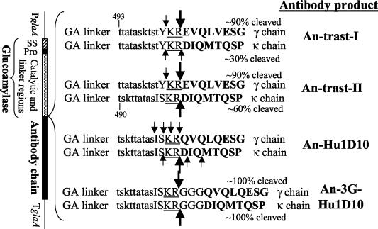
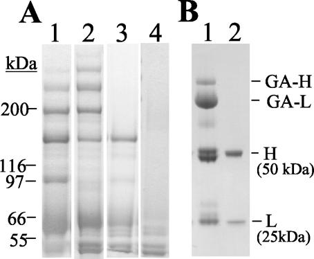
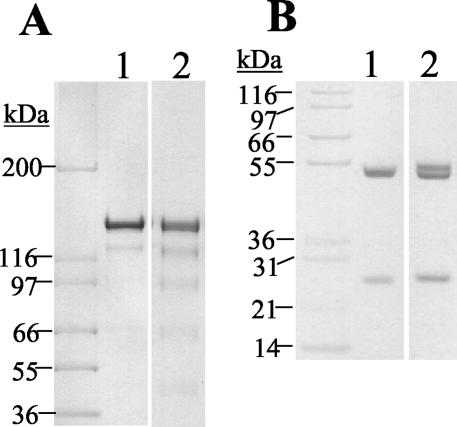
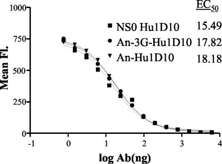
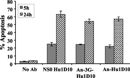
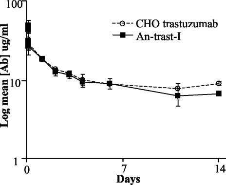
Two different humanized immunoglobulin G1(kappa) antibodies and an Fab' fragment were produced by Aspergillus niger. The antibodies were secreted into the culture supernatant. Both light and heavy chains were initially synthesized as fusion proteins with native glucoamylase. After antibody assembly, cleavage by A. niger KexB protease allowed the release of free antibody. Purification by hydrophobic charge induction chromatography proved effective at removing any antibody to which glucoamylase remained attached. Glycosylation at N297 in the Fc region of the heavy chain was observed, but this site was unoccupied on approximately 50% of the heavy chains. The glycan was of the high-mannose type, with some galactose present, and the size ranged from Hex(6)GlcNAc(2) to Hex(15)GlcNAc(2). An aglycosyl mutant form of antibody was also produced. No significant difference between the glycosylated antibody produced by Aspergillus and that produced by mammalian cell cultures was observed in tests for affinity, avidity, pharmacokinetics, or antibody-dependent cellular cytotoxicity function.


Figures

FIG. 1.
FIG. 1.
Schematic representation of the antibody light- and heavy-chain expression cassettes. The vertical bar on the left represents the expression cassette and the encoded proteins. PglaA, promoter region of the glaA gene; SS, glucoamylase signal sequence; Pro, glucoamylase proregion; TglaA, terminator region of the glaA gene. To the right are the designations of the various antibody products mentioned in the text. The amino acid sequence at the junction between the C terminus of the glucoamylase (GA) linker region (lowercase) and the N terminus of the antibody heavy (γ) or light (κ) chain (uppercase, bold) is shown for each antibody product. The KexB cleavage site (KR) is underlined. The amino acid numbers for mature glucoamylase are marked on the sequences. For each amino acid sequence, the approximate percentage of that fusion protein that was present in the cleaved form in culture supernatants is shown. For each amino acid sequence, the results of N-terminal sequence analysis of the cleaved form are shown; a large arrow indicates that >50% of the cleaved protein had that N terminus, and a small arrow indicates that <50% of the cleaved protein had that N terminus.
FIG. 2.
FIG. 2.
SDS-PAGE of secreted proteins stained with Coomassie brilliant blue. (A) Gel run under nonreducing conditions; each lane contained 10 μl of Aspergillus culture supernatant. Lane 1, An-trast-II; lane 2, An-Hu1D10; lane 3, An-3G-Hu1D10; lane 4, untransformed parent strain. (B) Antibody purified by protein A affinity chromatography. The gel was run under reducing conditions. Lane 1, An-trast-I; lane 2, commercially available trastuzumab. GA-H, glucoamylase- heavy-chain fusion protein; GA-L, glucoamylase- light-chain fusion protein; H, antibody heavy chain; L, antibody light chain.
FIG. 3.
FIG. 3.
SDS-PAGE of purified An-trast-I. (A) Gel run under nonreducing conditions. Lane 1, commercially available trastuzumab; lane 2, An-trast-I. (B) Gel run under reducing conditions. Lane 1, commercially available trastuzumab; lane 2, An-trast-I.
FIG. 4.
FIG. 4.
Competition binding assay. The amount of antibody (Ab) NS0-Hu1D10, An-3G-Hu1D10, or An-Hu1D10 that was mixed with 250 ng of FITC-labeled NS0-Hu1D10 is shown on the x axis. After incubation of the antibody mixtures with Raji cells, the resulting mean fluorescence (Fl) of the cell population was measured by flow cytometry and is shown on the y axis. EC50, 50% effective concentration.
FIG. 5.
FIG. 5.
Apoptosis assay. The percentages of Raji cell populations that underwent apoptosis after incubation for 5 or 24 h with NS0-Hu1D10, An-3G-Hu1D10, or An-Hu1D10 are shown. The data are averages from quadruplicate experiments, and the error bars indicate standard deviations.
FIG. 6.
FIG. 6.
Pharmacokinetics in rats. The mean concentrations of human IgG1(κ) antibody (Ab) in serum samples at various time points after injection of rats with either CHO cell-derived trastuzumab (four rats) or An-trast-I (three rats) are shown. Standard deviations are indicated by error bars.
FIG. 7.
FIG. 7.
ADCC assay. Raji cells and human PBMCs were mixed at a ratio of 1:40 and incubated with the amounts of NS0-Hu1D10, An-3G-Hu1D10, or An-Hu1D10 indicated on the x axes. The percentages of Raji cell populations that exhibited ADCC are shown on the y axes. (A) PBMCs obtained from one human donor. (B) PBMCs obtained from a second human donor. The data are averages from duplicate experiments.

References

    1. Ballance, D. J., F. P. Buxton, and G. Turner. 1983. Transformation of Aspergillus nidulans by the orotidine-5′-phosphate decarboxylase gene of Neurospora crassa. Biochem. Biophys. Res. Commun. 112:284-289. - PubMed
    1. Campbell, E. I., S. E. Unkles, J. A. Macro, C. van den Hondel, R. Contreras, and J. R. Kinghorn. 1989. Improved transformation efficiency of Aspergillus niger using the homologous niaD gene for nitrate reductase. Curr. Genet. 16:53-56. - PubMed
    1. Carter, P., R. F. Kelley, M. L. Rodrigues, B. Snedecor, M. Covarrubias, M. D. Velligan, W. L. T. Wong, A. M. Rowland, C. E. Kotts, M. E. Carver, M. Yang, J. H. Bourell, H. M. Shepard, and D. Henner. 1992. High level Escherichia coli expression and production of a bivalent humanized antibody fragment. Bio/Technology 10:163-167. - PubMed
    1. Carter, P., L. Presta, C. M. Gorman, J. B. B. Ridgway, D. Henner, W. L. T. Wong, A. M. Rowland, C. Kotts, M. E. Carver, and H. M. Shepard. 1992. Humanization of an anti-p185HER2 antibody for human cancer therapy. Proc. Natl. Acad. Sci. USA 89:4285-4289. - PMC - PubMed
    1. Dorai, H., B. M. Mueller, R. A. Reisfeld, and S. D. Gillies. 1991. Aglycosylated chimeric mouse/human IgG1 antibody retains some effector function. Hybridoma 10:211-217. - PubMed

LinkOut - more resources