

Spatial distribution and stability of the eight microbial species of the altered schaedler flora in the mouse gastrointestinal tract
- PMID: 15128534
- PMCID: PMC404395
- DOI: 10.1128/AEM.70.5.2791-2800.2004
Spatial distribution and stability of the eight microbial species of the altered schaedler flora in the mouse gastrointestinal tract
Abstract
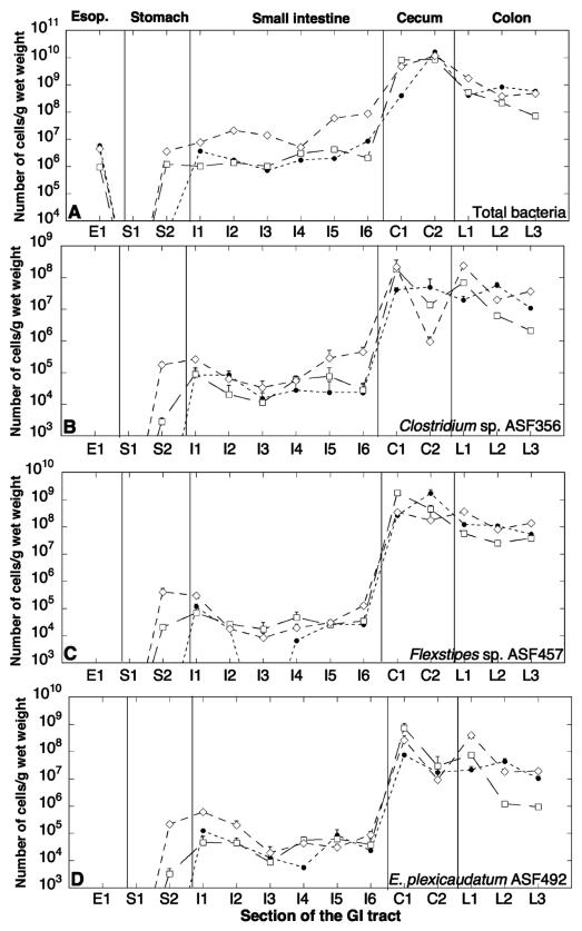
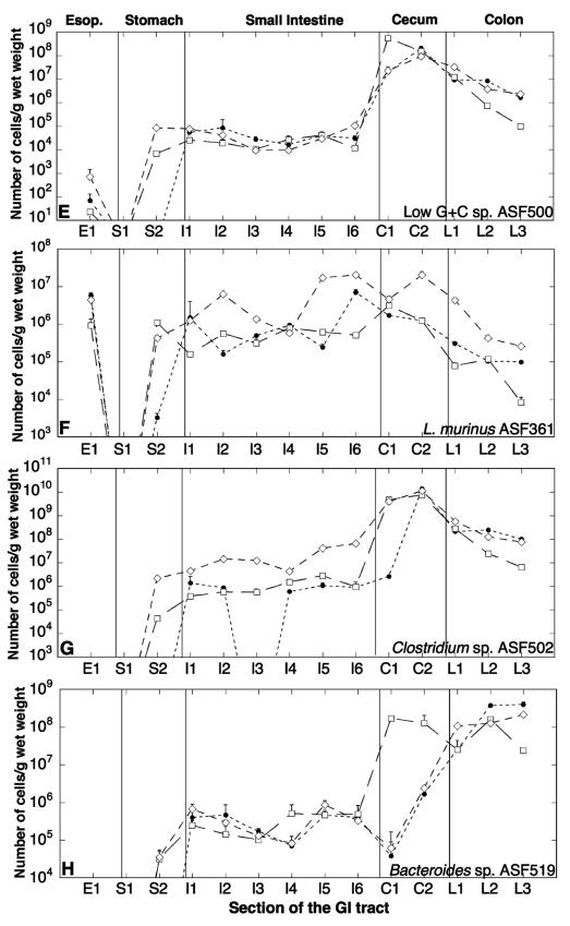
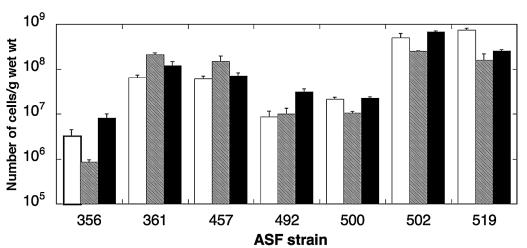
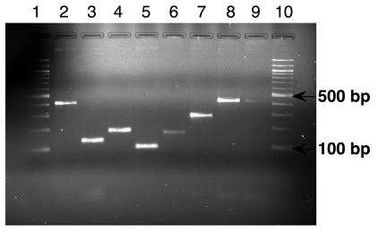
The overall complexity of the microbial communities in the gastrointestinal (GI) tracts of mammals has hindered observations of dynamics and interactions of individual bacterial populations. However, such information is crucial for understanding the diverse disease-causing and protective roles that gut microbiota play in their hosts. Here, we determine the spatial distribution, interanimal variation, and persistence of bacteria in the most complex defined-flora (gnotobiotic) model system to date, viz., mice colonized with the eight strains of the altered Schaedler flora (ASF). Quantitative PCR protocols based on the 16S rRNA sequence of each ASF strain were developed and optimized to specifically detect as few as 10 copies of each target. Total numbers of the ASF strains were determined in the different regions of the GI tracts of three C.B-17 SCID mice. Individual strain abundance was dependent on oxygen sensitivity, with microaerotolerant Lactobacillus murinus ASF361 present at 10(5) to 10(7) cells/g of tissue in the upper GI tract and obligate anaerobic ASF strains being predominant in the cecal and colonic flora at 10(8) to 10(10) cells/g of tissue. The variation between the three mice was small for most ASF strains, except for Clostridium sp. strain ASF502 and Bacteroides sp. strain ASF519 in the cecum. A comparison of the relative distribution of the ASF strains in feces and the colon indicated large differences, suggesting that fecal bacterial levels may provide a poor approximation of colonic bacterial levels. All ASF strains were detected by PCR in the feces of C57BL/6 restricted flora mice, which had been maintained in an isolator without sterile food, water, or bedding for several generations, providing evidence for the stability of these strains in the face of potential competition by bacteria introduced into the gut.
Figures
Similar articles
-
A real-time PCR assay for accurate quantification of the individual members of the Altered Schaedler Flora microbiota in gnotobiotic mice.J Microbiol Methods. 2017 Apr;135:52-62. doi: 10.1016/j.mimet.2017.02.003. Epub 2017 Feb 9. J Microbiol Methods. 2017. PMID: 28189782 Free PMC article.
-
Phylogeny of the defined murine microbiota: altered Schaedler flora.Appl Environ Microbiol. 1999 Aug;65(8):3287-92. doi: 10.1128/AEM.65.8.3287-3292.1999. Appl Environ Microbiol. 1999. PMID: 10427008 Free PMC article.
-
Charles River altered Schaedler flora (CRASF) remained stable for four years in a mouse colony housed in individually ventilated cages.Lab Anim. 2009 Oct;43(4):362-70. doi: 10.1258/la.2009.0080075. Epub 2009 Jun 17. Lab Anim. 2009. PMID: 19535393
-
The Altered Schaedler Flora: Continued Applications of a Defined Murine Microbial Community.ILAR J. 2015;56(2):169-78. doi: 10.1093/ilar/ilv012. ILAR J. 2015. PMID: 26323627 Free PMC article. Review.
-
Molecular ecological analysis of the gastrointestinal microbiota: a review.J Nutr. 2004 Feb;134(2):465-72. doi: 10.1093/jn/134.2.465. J Nutr. 2004. PMID: 14747690 Review.
Cited by
-
Helicobacter bilis Infection Alters Mucosal Bacteria and Modulates Colitis Development in Defined Microbiota Mice.Inflamm Bowel Dis. 2016 Nov;22(11):2571-2581. doi: 10.1097/MIB.0000000000000944. Inflamm Bowel Dis. 2016. PMID: 27755267 Free PMC article.
-
Colonization of the mouse upper gastrointestinal tract by lactobacillus murinus: a histological, immunocytochemical, and ultrastructural study.Curr Microbiol. 2013 Oct;67(4):395-8. doi: 10.1007/s00284-013-0367-9. Epub 2013 May 21. Curr Microbiol. 2013. PMID: 23689939
-
Spatial host-microbiome sequencing reveals niches in the mouse gut.Nat Biotechnol. 2024 Sep;42(9):1394-1403. doi: 10.1038/s41587-023-01988-1. Epub 2023 Nov 20. Nat Biotechnol. 2024. PMID: 37985876 Free PMC article.
-
Helicobacter bilis: bacterial provocateur orchestrates host immune responses to commensal flora in a model of inflammatory bowel disease.Gut. 2007 Jul;56(7):898-900. doi: 10.1136/gut.2006.115428. Gut. 2007. PMID: 17566023 Free PMC article. Review.
-
Pathogenic and non-pathogenic Escherichia coli colonization and host inflammatory response in a defined microbiota mouse model.Dis Model Mech. 2018 Nov 16;11(11):dmm035063. doi: 10.1242/dmm.035063. Dis Model Mech. 2018. PMID: 30275104 Free PMC article.
References
-
- Altschul, S. F., W. Gish, W. Miller, E. W. Myers, and D. J. Lipman. 1990. Basic local alignment search tool. J. Mol. Biol. 215:403-410. - PubMed
-
- Donaldson, R. M., and P. P. Toskes. 1989. The relation of enteric bacterial populations to gastrointestinal function and disease, p. 107-114. In J. S. Sleisenger (ed.), Gastrointestinal disease: pathophysiology, diagnosis, management. Saunders, Philadelphia, Pa.
Publication types
MeSH terms
Substances
Associated data
- Actions
Grants and funding
LinkOut - more resources
Full Text Sources
