

Comparative analysis of the conventional and novel pmo (particulate methane monooxygenase) operons from methylocystis strain SC2
- PMID: 15128567
- PMCID: PMC404415
- DOI: 10.1128/AEM.70.5.3055-3063.2004
Comparative analysis of the conventional and novel pmo (particulate methane monooxygenase) operons from methylocystis strain SC2
Abstract
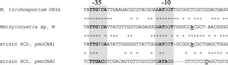
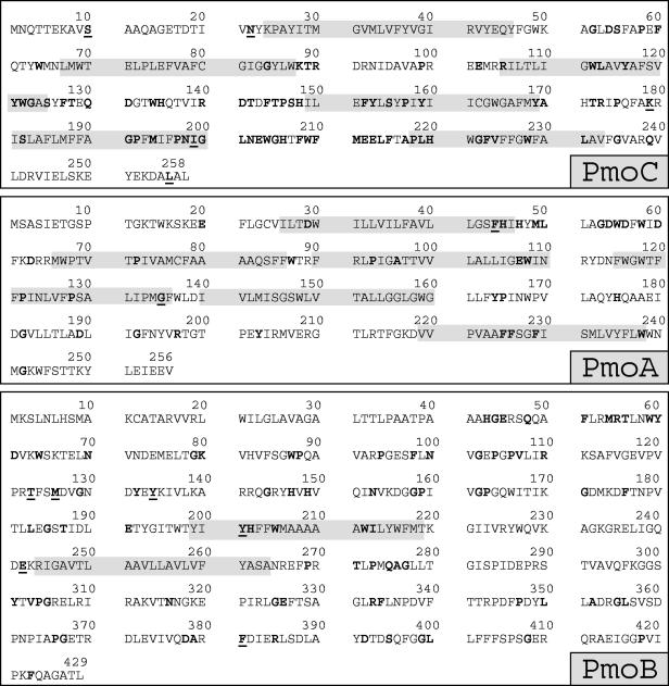
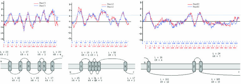
In addition to the conventional pmoA gene (pmoA1) encoding the active site polypeptide of particulate methane monooxygenase, a novel pmoA gene copy (pmoA2) is widely distributed among type II methanotrophs (methane-oxidizing bacteria [MOB]) (M. Tchawa Yimga, P. F. Dunfield, P. Ricke, J. Heyer, and W. Liesack, Appl. Environ. Microbiol. 69:5593-5602, 2003). Here we report that the pmoA1 and pmoA2 gene copies in the type II MOB Methylocystis strain SC2 are each part of a complete pmoCAB gene cluster (pmoCAB1, pmoCAB2). A bacterial artificial chromosome (BAC) library of strain SC2 genomic DNA was constructed, and BAC clones carrying either pmoCAB1 or pmoCAB2 were identified. Comparative sequence analysis showed that these two gene clusters exhibit low levels of identity at both the DNA level (67.4 to 70.9%) and the derived protein level (59.3 to 65.6%). In contrast, the secondary structures predicted for PmoCAB1 and PmoCAB2, as well as the derived transmembrane-spanning regions, are nearly identical. This suggests that PmoCAB2 is, like PmoCAB1, a highly hydrophobic, membrane-associated protein. A total of 190 of the 203 amino acid residues representing a highly conserved consensus sequence of the currently known PmoCAB1 and AmoCAB sequence types could be identified in PmoCAB2. The amoCAB gene cluster encodes ammonia monooxygenase and is evolutionarily related to pmoCAB. Analysis of a set of amino acid residues that allowed differentiation between conventional PmoA and AmoA provided further support for the hypothesis that pmoCAB2 encodes a functional equivalent of PmoCAB1. In experiments in which we used 5' rapid amplification of cDNA ends we identified transcriptional start sites 320 and 177 bp upstream of pmoC1 and pmoC2, respectively. Immediately upstream of the transcriptional start sites of both pmoCAB1 and pmoCAB2, sequence motifs similar to Escherichia coli sigma(70) promoters were identified.
Figures
Similar articles
-
Two isozymes of particulate methane monooxygenase with different methane oxidation kinetics are found in Methylocystis sp. strain SC2.Proc Natl Acad Sci U S A. 2008 Jul 22;105(29):10203-8. doi: 10.1073/pnas.0702643105. Epub 2008 Jul 15. Proc Natl Acad Sci U S A. 2008. PMID: 18632585 Free PMC article.
-
Wide distribution of a novel pmoA-like gene copy among type II methanotrophs, and its expression in Methylocystis strain SC2.Appl Environ Microbiol. 2003 Sep;69(9):5593-602. doi: 10.1128/AEM.69.9.5593-5602.2003. Appl Environ Microbiol. 2003. PMID: 12957949 Free PMC article.
-
Molecular analysis of the pmo (particulate methane monooxygenase) operons from two type II methanotrophs.Appl Environ Microbiol. 2000 Mar;66(3):966-75. doi: 10.1128/AEM.66.3.966-975.2000. Appl Environ Microbiol. 2000. PMID: 10698759 Free PMC article.
-
Ammonium induces differential expression of methane and nitrogen metabolism-related genes in Methylocystis sp. strain SC2.Environ Microbiol. 2014 Oct;16(10):3115-27. doi: 10.1111/1462-2920.12367. Epub 2014 Feb 18. Environ Microbiol. 2014. PMID: 24373058
-
Role of multiple gene copies in particulate methane monooxygenase activity in the methane-oxidizing bacterium Methylococcus capsulatus Bath.Microbiology (Reading). 1999 May;145 ( Pt 5):1235-1244. doi: 10.1099/13500872-145-5-1235. Microbiology (Reading). 1999. PMID: 10376840
Cited by
-
Genomic Analysis of the Yet-Uncultured Binatota Reveals Broad Methylotrophic, Alkane-Degradation, and Pigment Production Capacities.mBio. 2021 May 18;12(3):e00985-21. doi: 10.1128/mBio.00985-21. mBio. 2021. PMID: 34006650 Free PMC article.
-
Microbial CH(4) and N(2)O Consumption in Acidic Wetlands.Front Microbiol. 2012 Mar 2;3:78. doi: 10.3389/fmicb.2012.00078. eCollection 2012. Front Microbiol. 2012. PMID: 22403579 Free PMC article.
-
Methane-oxidizing bacteria in a California upland grassland soil: diversity and response to simulated global change.Appl Environ Microbiol. 2005 May;71(5):2642-52. doi: 10.1128/AEM.71.5.2642-2652.2005. Appl Environ Microbiol. 2005. PMID: 15870356 Free PMC article.
-
Two isozymes of particulate methane monooxygenase with different methane oxidation kinetics are found in Methylocystis sp. strain SC2.Proc Natl Acad Sci U S A. 2008 Jul 22;105(29):10203-8. doi: 10.1073/pnas.0702643105. Epub 2008 Jul 15. Proc Natl Acad Sci U S A. 2008. PMID: 18632585 Free PMC article.
-
Complete genome sequence of Methylocystis sp. strain SC2, an aerobic methanotroph with high-affinity methane oxidation potential.J Bacteriol. 2012 Nov;194(21):6008-9. doi: 10.1128/JB.01446-12. J Bacteriol. 2012. PMID: 23045511 Free PMC article.
References
-
- Altschul, S. F., W. Gish, W. Miller, E. W. Myers, and D. J. Lipman. 1990. Basic local alignment search tool. J. Mol. Biol. 215:403-410. - PubMed
-
- Choi, D. W., R. C. Kunz, E. S. Boyd, J. D. Semrau, W. E. Antholine, J. I. Han, J. A. Zahn, J. M. Boyd, A. M. de la Mora, and A. A. DiSpirito. 2003. The membrane-associated methane monooxygenase (pMMO) and pMMO-NADH:quinone oxidoreductase complex from Methylococcus capsulatus Bath. J. Bacteriol. 185:5755-5764. - PMC - PubMed
Publication types
MeSH terms
Substances
Associated data
- Actions
- Actions
LinkOut - more resources
Full Text Sources
