. 2004 Sep;71(3):828-36.
doi: 10.1095/biolreprod.104.030569. Epub 2004 May 5.

Gonadotropin-releasing hormone regulates expression of the DNA damage repair gene, Fanconi anemia A, in pituitary gonadotroph cells

Affiliations

Gonadotropin-releasing hormone regulates expression of the DNA damage repair gene, Fanconi anemia A, in pituitary gonadotroph cells

Rachel Larder et al. Biol Reprod. 2004 Sep.

Abstract

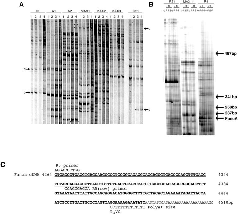
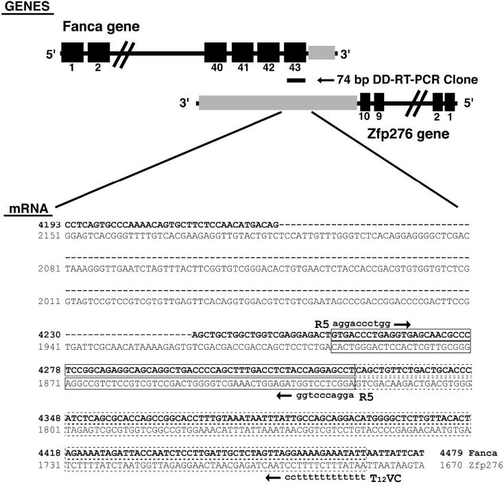
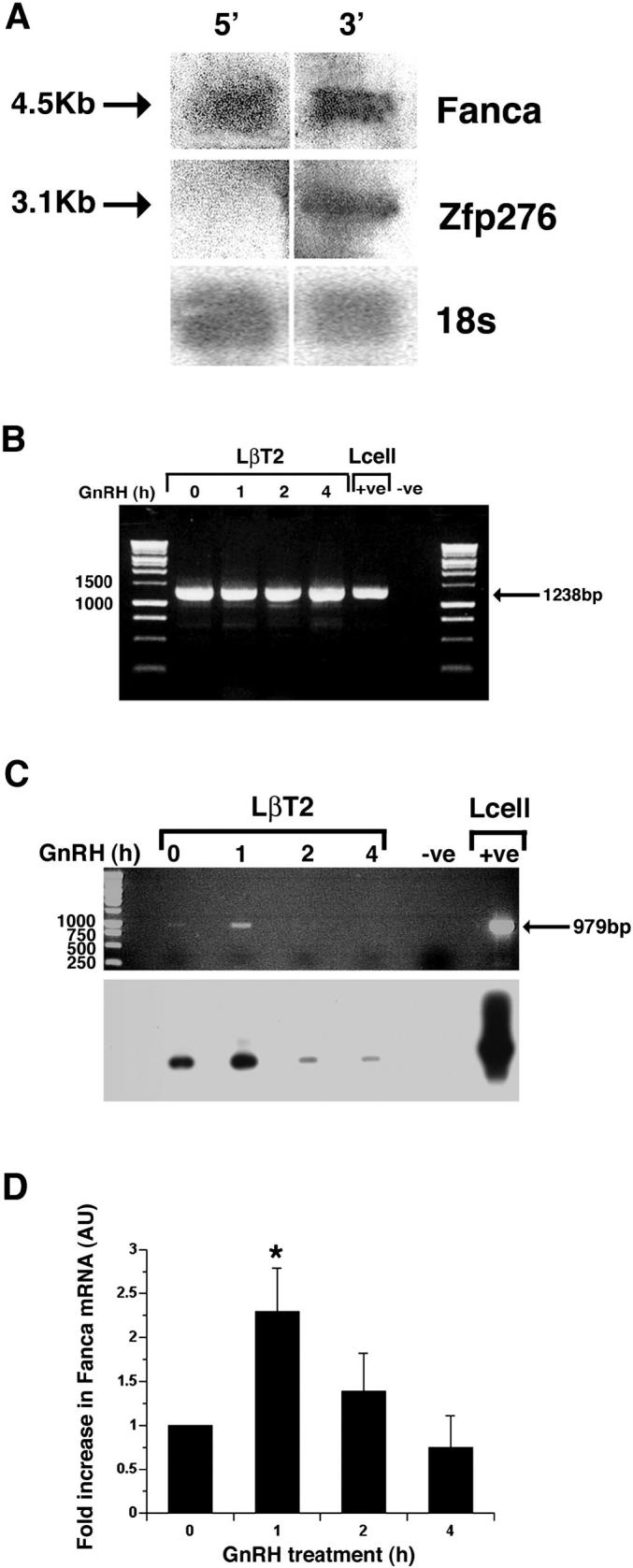
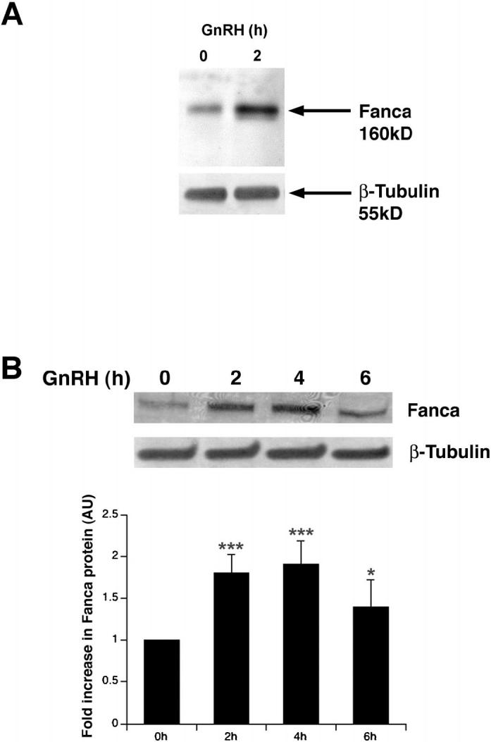
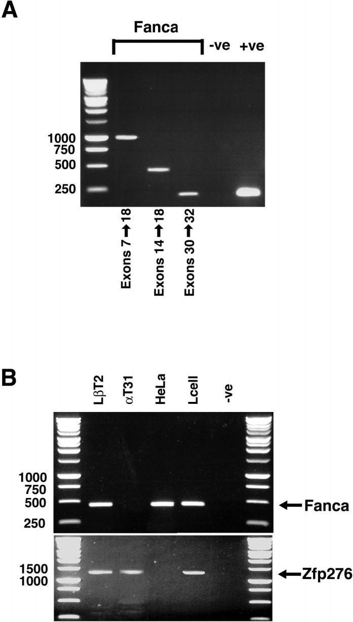
Gonadal function is critically dependant on regulated secretion of the gonadotropin hormones from anterior pituitary gonadotroph cells. Gonadotropin biosynthesis and release is triggered by the binding of hypothalamic GnRH to GnRH receptor expressed on the gonadotroph cell surface. The repertoire of regulatory molecules involved in this process are still being defined. We used the mouse L beta T2 gonadotroph cell line, which expresses both gonadotropin hormones, as a model to investigate GnRH regulation of gene expression and differential display reverse transcription-polymerase chain reaction (RT-PCR) to identify and isolate hormonally induced changes. This approach identified Fanconi anemia a (Fanca), a gene implicated in DNA damage repair, as a differentially expressed transcript. Mutations in Fanca account for the majority of cases of Fanconi anemia (FA), a recessively inherited disease identified by congenital defects, bone marrow failure, infertility, and cancer susceptibility. We confirmed expression and hormonal regulation of Fanca mRNA by quantitative RT-PCR, which showed that GnRH induced a rapid, transient increase in Fanca mRNA. Fanca protein was also acutely upregulated after GnRH treatment of L beta T2 cells. In addition, Fanca gene expression was confined to mature pituitary gonadotrophs and adult mouse pituitary and was not expressed in the immature alpha T3-1 gonadotroph cell line. Thus, this study extends the expression profile of Fanca into a highly specialized endocrine cell and demonstrates hormonal regulation of expression of the Fanca locus. We suggest that this regulatory mechanism may have a crucial role in the GnRH-response mechanism of mature gonadotrophs and perhaps the etiology of FA.


Figures

FIG. 1
FIG. 1
Differential display RT-PCR analysis of GnRH-regulated transcripts isolated from LβT2 cells. A) Matrigel basement membrane was excluded as an inducer of gene expression in LβT2 gonadotroph cells while GnRH was shown to upregulate transcripts. Cells were grown without Matrigel (1) or were cultured on Matrigel (2, 3, and 4) and treated with 3 (3) or 6 (4) 15-min pulses of GnRH with an interpulse interval of 75 min. RNA was extracted and subjected to differential display (DD) RT-PCR using primers stated. Matrigel did not alter transcript expression (arrows a and b), while numerous transcripts altered after GnRH treatment (arrows c and d). B) In two separate experiments, LβT2 cells were left untreated (−G) or were treated with 1 × 15-min pulse of GnRH (+G), RNA was harvested (−G, 1, 2, and 4 h) and subjected to DD-RT-PCR. First-strand cDNA was generated using the T12VC downstream primer, then amplified by PCR using primers R21, MAX1, or R5. Arrow denotes location of Fanca. The location of DNA size markers, indicated as bp, are also shown. C) Bioinformatic line-up depicting the region of homology, shown underlined, between the cloned Fanca DD-RT-PCR product and mouse Fanca cDNA nucleotide sequence, and the likely internal R5 priming site. The location of DD-RT-PCR primers R5 and T12VC that generated the original 216-bp DD-RT-PCR product are also indicated.
FIG. 2
FIG. 2
Schematic representation of the Fanca locus showing the location of the differential display RT-PCR clone. Exons 40-43 of Fanca, shown as black boxes, overlap with the 3′ untranslated region (UTR) of Zfp276, shown in grey. The location of the cloned 74-bp differential display (DD) RT-PCR clone is indicated. Beneath the schematic, the regions of nucleotide identity between Fanca mRNA and Zfp276 mRNA have been aligned, Fanca mRNA sequence is shown in bold type, and spliced intronic sequence has been omitted and replaced with a dashed line, with Zfp276 mRNA in normal typeface. The amplified 74-bp DD-RT-PCR clone has been boxed with a solid line, and the original 216-bp DD-RT-PCR clone is indicated as an extension of the boxed region with dashed lines. The amplification primers R5 and T12VC are also shown on the corresponding Fanca sequence. No suitable priming sites were identified by bioinformatic analysis for the T12VC-anchored primer on the Zfp276 3′ UTR sequence.
FIG. 3
FIG. 3
Characterization and quantification of Fanca and Zfp276 mRNA after GnRH treatment. A) Confirmation of expression of full-length 4.5-kb Fanca and 3.1-kb Zfp276 mRNA transcripts in LβT2 cells. Total RNA from LβT2 cells was fractionated on a formaldehyde gel, Northern blotted, and probed with radiolabeled probes corresponding to either the 5′ region (exons 1-11) or 3′ region (exons 42-43) of Fanca. The blot was then stripped and reprobed with an 18s probe. Specific Fanca, Zfp276, and 18s bands are indicated by arrows. B) Semiquantitative RT-PCR analysis of Zfp276 expression. LβT2 cells were left untreated (0) or treated with 1 × 15-min pulse of GnRH, then harvested 1, 2, and 4 h later, RNA was extracted, and first-strand cDNA made. PCR was performed to amplify full-length Zfp276 (1238 bp) and the products were visualized on an ethidium bromide-stained agarose gel. One specific 1238-bp PCR product was visible at all time points. L-cell cDNA was included as a positive control. An arrow denotes the 1238-bp PCR product, and DNA size markers are labeled. C) Semiquantitative RT-PCR analysis was performed for Fanca expression in LβT2 cells that were left untreated (0) or treated with 1 pulse of GnRH; harvested 1, 2, and 4 h later; RNA extracted, and cDNA made. Ethidium bromide staining identified one PCR product amplified from mRNA harvested from the 1-h time point. L-cell cDNA was included as a positive control. An arrow denotes the 979-bp PCR product, and size markers are labeled. Southern blotting analysis of the agarose gel with a 5′ Fanca probe confirmed that the Fanca PCR product was specific and present at all time points. D) LightCycler quantitative RT-PCR analysis of Fanca mRNA extracted from LβT2 cells either left untreated or treated with GnRH and harvested 1, 2, and 4 h later detected a consistent 2-fold increase in Fanca mRNA harvested 1 h after treatment. Results are shown as arbitrary units (AU) of Fanca mRNA normalized against the levels of internal control B-2-microglobulin (B2m) mRNA. This experiment was performed in duplicate and repeated three times. *P < 0.05 was determined as being significant by ANOVA one-way analysis of variance.
FIG. 4
FIG. 4
Western blotting analysis of Fanca protein. A) Western blotting analysis of protein extracts from untreated, 0, and extracts harvested 2 h after GnRH treatment identified Fanca protein. Cellular protein was fractionated, transferred to PVDF, and probed with anti-mouse Fanca antisera before being stripped and reprobed with anti-mouse β-tubulin. Arrows indicate the 160-kDa Fanca and 55-kDa β-tubulin proteins. B) Western blotting analysis of whole-cell protein extracts from untreated (0) and hormone-treated cells harvested 2, 4, and 6 h later. Blots were probed as above in A. ANOVA one-way analysis of variance determined that the increases in Fanca protein levels after hormone treatment were significant: ***P < 0.001; ***P < 0.001; *P < 0.05.
FIG. 5
FIG. 5
Fanca is expressed in adult mouse pituitary. A) RT-PCR analysis of RNA extracted from adult mouse pituitary was performed using primers that amplified exons 7-18, exons 14-18, and exons 30-32 of mouse Fanca. Ethidium bromide staining identified PCR products for all regions amplified. A control GAPDH PCR product was also amplified, confirming the integrity of the mouse pituitary cDNA. B) RNA was extracted from LβT2, αT3-1, HeLa, and L cells; reverse transcribed; and first-strand cDNA was analyzed by PCR for expression of Fanca. The PCR primers corresponded to exons 7-18 of mouse Fanca cDNA and ethidium bromide staining identified a 450-bp product in LβT2, HeLa, and L cells. No Fanca expression was detectable when amplifying αT3-1 cDNA. In this experiment, expression of Zfp276 was used as an internal control to check PCR conditions and RNA integrity. A specific 1238-bp PCR product, corresponding to Zfp276, was amplified from LβT2, αT3-1, and L cell first-strand cDNA.

References

    1. Stanislaus D, Pinter JH, Janovick JA, Conn PM. Mechanisms mediating multiple physiological responses to gonadotropin-releasing hormone. Mol Cell Endocrinol. 1998;144:1–10. - PubMed
    1. Brown P, McNeilly AS. Transcriptional regulation of pituitary gonadotrophin subunit genes. Rev Reprod. 1999;4:117–124. - PubMed
    1. Shacham S, Harris D, Ben-Shlomo H, Cohen I, Bonfil D, Przedecki F, Lewy H, Ashkenazi IE, Seger R, Naor Z. Mechanism of GnRH receptor signaling on gonadotropin release and gene expression in pituitary gonadotrophs. Vitam Horm. 2001;63:63–90. - PubMed
    1. Charlton HM, Halpin DM, Iddon C, Rosie R, Levy G, McDowell I, Megson A, Morris J, Bramwell A, Speight A, Ward B, Broadhead J, Davey-Smith G, Fink G. The effects of daily administration of single and multiple injections of gonadotropin releasing hormone on pituitary and gonadal function in the hypogonadal (HPG) mouse. Endocrinology. 1983;113:535–544. - PubMed
    1. Brown P, McNeilly J, Evans J, Crawford G, Walker M, Christian H, McNeilly A. Manipulating the in vivo mRNA expression profile of FSHβ to resemble that of LHβ does not promote a concomitant increase in intracellular storage of follicle-stimulating hormone. J Neuroendocrinol. 2001;13:50–62. - PubMed

Publication types

MeSH terms

Substances