. 2004 May 15;172(10):6057-64.
doi: 10.4049/jimmunol.172.10.6057.

T cells associated with tumor regression recognize frameshifted products of the CDKN2A tumor suppressor gene locus and a mutated HLA class I gene product

Affiliations

T cells associated with tumor regression recognize frameshifted products of the CDKN2A tumor suppressor gene locus and a mutated HLA class I gene product

Jianping Huang et al. J Immunol. .

Abstract

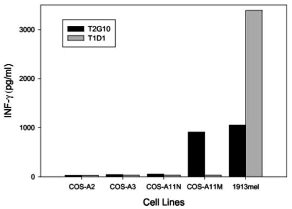
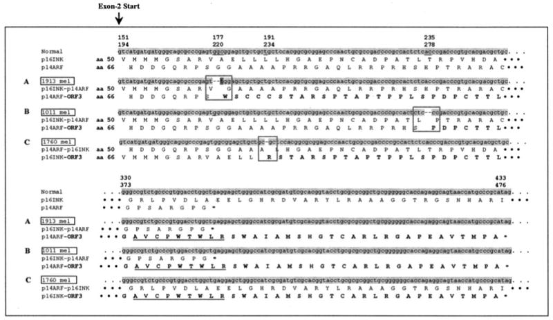
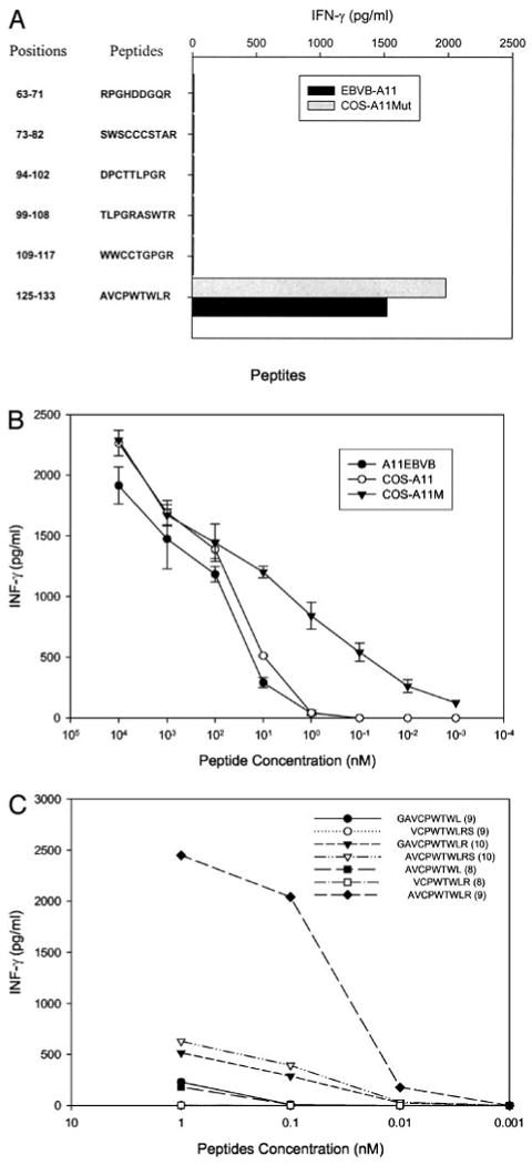
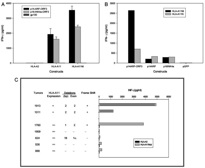
The dramatic tumor regression observed following adoptive T cell transfer in some patients has led to attempts to identify novel Ags to understand the nature of these responses. Nearly complete regression of multiple metastatic melanoma lesions was observed in patient 1913 following adoptive transfer of autologous tumor-infiltrating lymphocytes. The autologous 1913 melanoma cell line expressed a mutated HLA-A11 class I gene product that was recognized by the bulk tumor-infiltrating lymphocytes as well as a dominant T cell clone derived from this line. A second dominant T cell clone, T1D1, did not recognize the mutated HLA-A11 product, but recognized an allogeneic melanoma cell line that shared expression of HLA-A11 with the parental tumor cell line. Screening of an autologous melanoma cDNA library with clone T1D1 T cells in a cell line expressing the mutated HLA-A11 gene product resulted in the isolation of a p14ARF transcript containing a 2-bp deletion in exon 2. The T cell epitope recognized by T1D1, which was encoded within the frameshifted region of the deleted p14ARF transcript, was also generated from frameshifted p14ARF or p16INK4a transcripts that were isolated from two additional melanoma cell lines. The results of monitoring studies indicated that T cell clones reactive with the mutated HLA-A11 gene product and the mutated p14ARF product were highly represented in the peripheral blood of patient 1913 1 wk following adoptive transfer, indicating that they may have played a role in the nearly complete tumor regression that was observed following this treatment.


Figures

FIGURE 1
FIGURE 1
Recognition of HLA-A11Mut by clone T2G10. COS-7 cells were transfected for 24 h with HLA-A11Mut, HLA-A11N, HLA-A3, or HLA-A2. A coculture was then conducted with either clone T2G10 or T1D1, and the release of IFN-γ was measured 18 h later. The autologous 1913 mel cell line was also included in the coculture panel as a positive control.
FIGURE 2
FIGURE 2
Sequences of frameshifted p14-ARF and p16-INK4a gene products expressed by melanoma cells. The partial nucleotide and amino acid sequences of the p14-ARF and p16-INK4a transcripts that were cloned from the three melanoma cell lines 1913 mel (A), 1011 mel (B), and 1760 mel (C) are presented in this figure. Deleted nucleotides are indicated by dashes, and the point mutation is highlighted by the dark gray. Amino acid sequences encoded within ORF3 are noted in bold. The first two amino acid sequences represent those that are encoded by the normal p16INK4a and p14ARF transcript, and the lower set of amino acid sequences represent those that are encoded by the frameshifted transcripts. The amino acids that flank the mutation sites are boxed, and the sequence of the T cell epitope is underlined.
FIGURE 3
FIGURE 3
Identification and titration of the p16INK4a/p14ARF peptide recognized by clone T1D1. A, Peptide screening. Six peptides were synthesized and incubated for 2 h with A11-EBVB and COS-A11Mut cells at a concentration of 100 nM. Target cells were then cocultured with clone T1D1, and IFN-γ release was measured 18 h later. B, Peptide titration. Either HLA-A11-expressing EBVB, COS-A11N, or COS-A11Mut cells were loaded with the indicated concentrations of the AVCPWTWLR peptide for 2 h; clone T1D1 cells were added; and IFN-γ release was measured 18 h later. C, Testing of peptide variants. Variants of the peptide AVCPWTWLR either lacking or containing an additional N- or C-terminal residue were incubated with COS-A11Mut cells at indicated concentrations for 2 h. Clone T1D1 T cells were then added, and IFN-γ release was measured 18 h later.
FIGURE 4
FIGURE 4
T1D1/CL131 recognition of melanomas and transfected cells with frameshifted and normal p16INK4a and p14ARF gene products. A, CL131 recognition of cells transfected with constructs encoding p14ARF-ORF3 or p16INK4a-ORF3. COS-A2, COS-A11N, and COS-A11Mut cells were transiently transfected with constructs encoding p14ARF-ORF3, p16INK4a-ORF3, or gp100; 24 h later, clone CL131 T cells were added; and IFN-γ release was measured 18 h later. B, Relative recognition of target cells transfected with constructs encoding p14ARF-ORF3, normal p14ARF, or normal p16INK4a by T1D1. The COS-A11N and COS-A11Mut lines were transfected with the indicated constructs; 24 h later, clone T1D1 T cells were added; and IFN-γ release was measured 18 h later. C, Recognition of melanoma cell lines that express frameshifted or nonframeshifted p14ARF or p16INK4a transcripts by T1D1 cells. The 1913, 1011, and 1760 melanoma cell lines, which express frameshifted transcripts, as well as four additional melanoma cell lines, 1909, 624, 526, and 888, which did not express frameshifted transcripts, were incubated with T1D1 T cells, and IFN-γ release was measured 18 h later. The 1760, 1909, 624, 526, and 888 mel cell lines, which do not naturally express HLA-A11, were transduced with recombinant retroviral constructs that encode either HLA-A2 or the HLA-A11mut gene isolated from 1913 mel, as indicated.

References

    1. Robbins PF, Wang RF, Rosenberg SA. Tumor antigens recognized by cytotoxic lymphocytes. In: Sitkovsky MV, Henkart PA, editors. Cytotoxic Cells: Basic Mechanisms and Medical Applications. Lippincott Williams & Wilkins; Philadelphia: 2000. p. 363.
    1. Ikeda H, Lethe B, Lehmann F, van Baren N, Baurain JF, de Smet C, Chambost H, Vitale M, Moretta A, Boon T, Coulie PG. Characterization of an antigen that is recognized on a melanoma showing partial HLA loss by CTL expressing an NK inhibitory receptor. Immunity. 1997;6:199. - PubMed
    1. Hanada K, Perry-Lalley DM, Ohnmacht GA, Bettinotti MP, Yang JC. Identification of fibroblast growth factor-5 as an overexpressed antigen in multiple human adenocarcinomas. Cancer Res. 2001;61:5511. - PubMed
    1. Wolfel T, Hauer M, Schneider J, Serrano M, Wolfel C, Klehmann-Hieb E, De Plaen E, Hankeln T, Meyer zum Buschenfelde KH, Beach D. A p16INK4a-insensitive CDK4 mutant targeted by cytolytic T lymphocytes in a human melanoma. Science. 1995;269:1281. - PubMed
    1. Mandruzzato S, Brasseur F, Andry G, Boon T, van der Bruggen P. A CASP-8 mutation recognized by cytolytic T lymphocytes on a human head and neck carcinoma. J Exp Med. 1997;186:785. - PMC - PubMed

MeSH terms