. 2004 May 15;172(10):6087-92.
doi: 10.4049/jimmunol.172.10.6087.

Glycoprotein 96 can chaperone both MHC class I- and class II-restricted epitopes for in vivo presentation, but selectively primes CD8+ T cell effector function

Affiliations

Glycoprotein 96 can chaperone both MHC class I- and class II-restricted epitopes for in vivo presentation, but selectively primes CD8+ T cell effector function

Amy D H Doody et al. J Immunol. .

Abstract

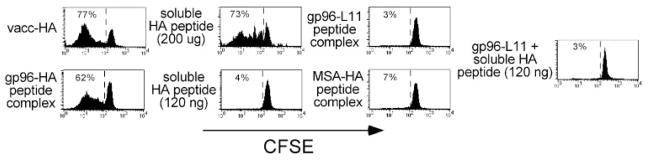
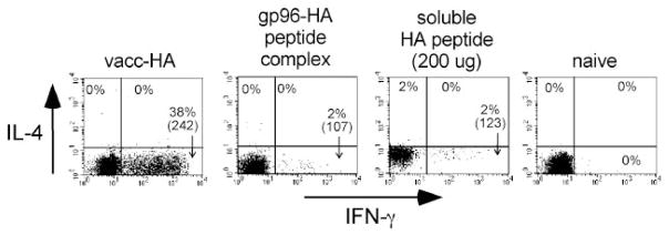
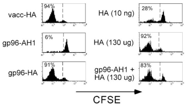
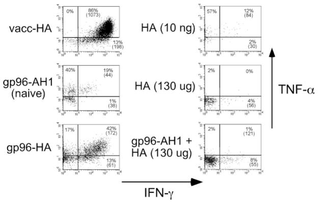
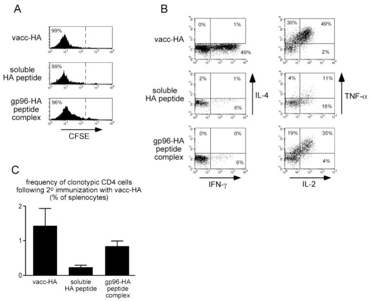
The ability of mature T lymphocytes to develop effector capacity after encounter with cognate Ag is generally dependent upon inflammatory signals associated with infection that induce dendritic cell activation/maturation. These inflammatory signals can derive directly from pathogens or can be expressed by host cells in response to infection. Heat shock proteins (HSPs) are a class of host-derived inflammatory mediators that perform the dual function of both chaperoning MHC class I-restricted epitopes into the cross-presentation pathway of DCs and inducing the activation/maturation of these DCs to allow priming of cognate CD8(+) T cell effector responses. Although the ability of HSPs to elicit effector CD8 cell responses has been well established, their potential to prime CD4 cell effector responses has been relatively unexplored. In the current study we compared the ability of the endoplasmic reticulum-resident HSP gp96 to prime CD4 vs CD8 cells using TCR transgenic adoptive transfer systems and soluble gp96-peptide complexes. As expected, gp96 facilitated the cross-presentation of a class I-restricted peptide and priming of effector function in cognate CD8 cells. Interestingly, gp96 also facilitated the in vivo presentation of a class II-restricted peptide; however, the resulting CD4 cell response did not involve the development of effector function. Taken together, these data suggest that gp96 is an inflammatory mediator that selectively primes CD8 cell effector function.


Figures

FIGURE 1
FIGURE 1
gp96 can facilitate in vivo presentation of MHC class II-restricted epitopes. Naive Thy1.1+ CFSE-labeled clonotypic CD4 cells were adoptively transferred into Thy1.2+ NT recipients that were simultaneously immunized intradermally with vacc-HA, 200 μg or 120 ng of soluble HA peptide, 70 μg of gp96-HA peptide complexes, 70 μg of gp96-L11 peptide complexes, 70 μg of MSA-HA peptide complexes, or 70 μg of gp96-L11 complexes and 120 ng of soluble HA peptide. Four days later, clonotypic CD4 cells (Thy1.1+) were recovered from the draining LNs and analyzed for their proliferative response (i.e., CFSE dilution). Each histogram is representative of three separate replicates.
FIGURE 2
FIGURE 2
gp96 cannot elicit CD4 cell effector function. Clonotypic CD4 cells recovered from recipients described in Fig. 1 that had undergone significant proliferation were restimulated in vitro with HA peptide-pulsed APCs and subsequently stained for intracellular IFN-γ and IL-4 expression. The naive group derived from the undivided cells that had been exposed to gp96-L11 peptide complexes. FACS plots show IFN-γ vs IL-4 expression, with the percentage of clonotypic CD4 cells expressing each cytokine (as well as the level of IFN-γ expression (mean fluorescence intensity) in parentheses) shown for each quadrant. Each plot is representative of three separate replicates. MiCK-2 cells (BD PharMingen) were used to verify that staining reactions contained a saturating concentration of anti-IL-4 mAb (data not shown).
FIGURE 3
FIGURE 3
gp96 can facilitate in vivo presentation of MHC class I-restricted epitopes. Naive Thy1.1+ CFSE-labeled clonotypic CD8 cells were adoptively transferred into Thy1.2+ NT recipients that were simultaneously immunized intradermally with vacc-HA, 130 μg or 10 ng of soluble HA peptide, 7 μg of gp96-HA peptide complexes, 7 μg of gp96-AH1 peptide complexes, or 7 μg of gp96-AH1 complexes and 130 μg of soluble HA peptide. Four days later, clonotypic CD8 cells (Thy1.1+) were recovered from the draining LNs and analyzed for their proliferation response (i.e., CFSE dilution). Each histogram is representative of three separate replicates, except for the 7 μg of gp96-AH1 complexes and 130 μg of soluble HA peptide group, which is representative of two replicates.
FIGURE 4
FIGURE 4
gp96 can elicit CD8 cell effector function. Clonotypic CD8 cells recovered from the recipients described in Fig. 3 were restimulated in vitro with HA peptide-pulsed APCs and subsequently stained for intracellular IFN-γ and TNF-α expression. The naive group derived from the undivided cells that had been exposed to gp96-AH1 peptide complexes. Representative FACS plots of IFN-γ vs TNF-α expression, with the percentage of clonotypic CD8 cells expressing each cytokine (as well as the level of IFN-γ expression (mean fluorescence intensity) in parentheses) shown for each quadrant.
FIGURE 5
FIGURE 5
gp96 does not induce CD4 cell tolerance. Naive clonotypic CD4 cell adoptive transfer recipients were immunized intradermally with 200 μg of soluble HA peptide or 52 μg of gp96-HA peptide complex on 4 consecutive days or once with vacc-HA. Clonotypic CD4 cells were recovered from the draining LNs on day 5 and analyzed for CFSE dilution (gating on Thy1.1+ and 6.5 (anti-clonotypic TCR) + cells); A) and intracellular cytokine expression after in vitro restimulation with HA peptide-pulsed APCs (gating on Thy1.1+ and CFSEdim cells; B). Each histogram is representative of three separate replicates. C, Clonotypic CD4 cells (3.5 × 10⁴) recovered from each of the primary adoptive transfer recipients described in A and B were retransferred into vacc-HA-immunized secondary recipients, and their frequencies were measured 6 days later after recovery from spleens. Data are expressed as the mean ± SEM (n = 3 for each group).

References

    1. Matzinger P. Tolerance, danger, and the extended family. Annu Rev Immunol. 1994;12:991. - PubMed
    1. Hawiger D, Inaba K, Dorsett Y, Guo M, Mahnke K, Rivera M, Ravetch JV, Steinman RM, Nussenzweig MC. Dendritic cells induce peripheral T cell unresponsiveness under steady state conditions in vivo. J Exp Med. 2001;194:769. - PMC - PubMed
    1. Jenkins MK, Khoruts A, Ingulli E, Mueller DL, McSorley SJ, Reinhardt RL, Itano A, Pape KA. In vivo activation of antigen-specific CD4 T cells. Annu Rev Immunol. 2001;19:23. - PubMed
    1. Janeway CA, Jr, Medzhitov R. Innate immune recognition. Annu Rev Immunol. 2002;20:197. - PubMed
    1. Vella AT, McCormack JE, Linsley PS, Kappler JW, Marrack P. Lipopolysaccharide interferes with the induction of peripheral T cell death. Immunity. 1995;2:261. - PubMed

Publication types

MeSH terms

Substances