. 2004 May 5;24(18):4313-23.
doi: 10.1523/JNEUROSCI.3303-03.2004.

Four classes of intercellular channels between glial cells in the CNS

Affiliations

Four classes of intercellular channels between glial cells in the CNS

Bruce M Altevogt et al. J Neurosci. .

Abstract

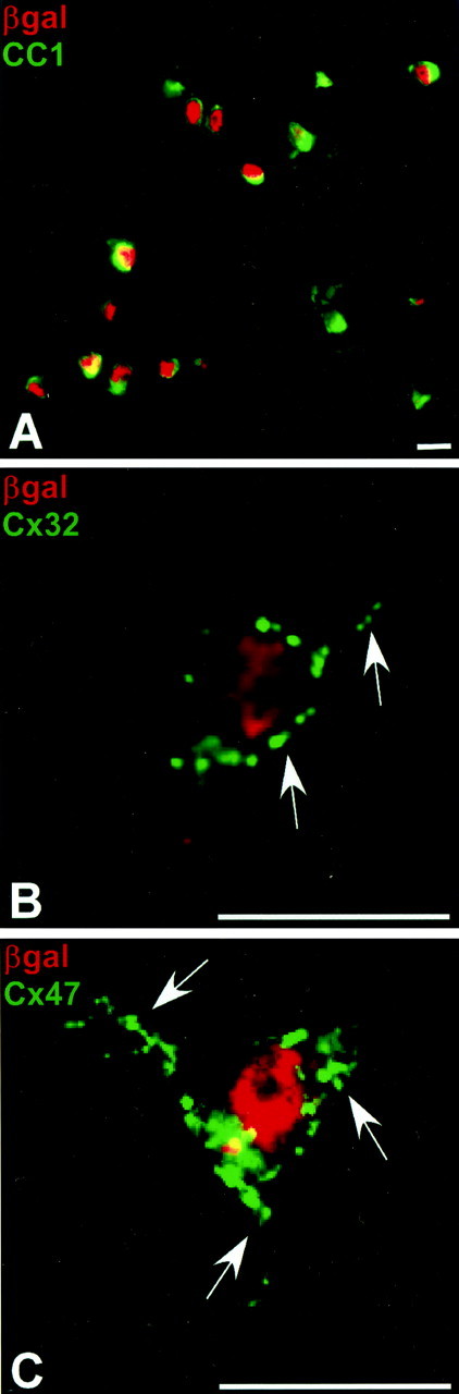
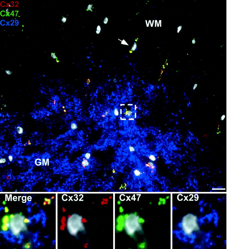
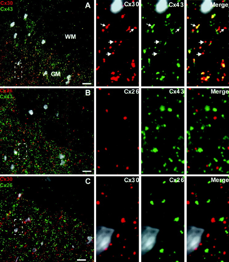
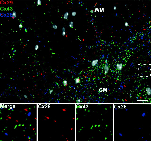
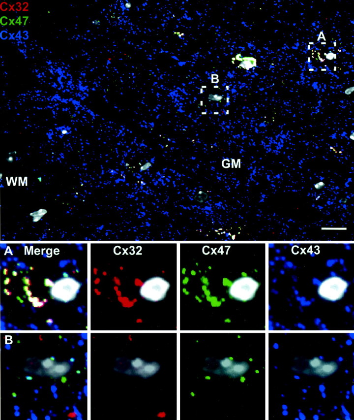
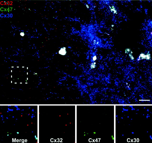
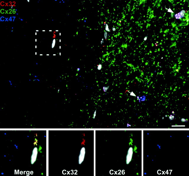
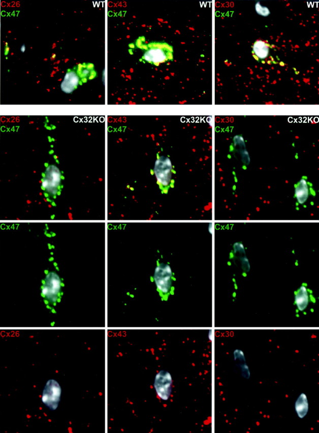
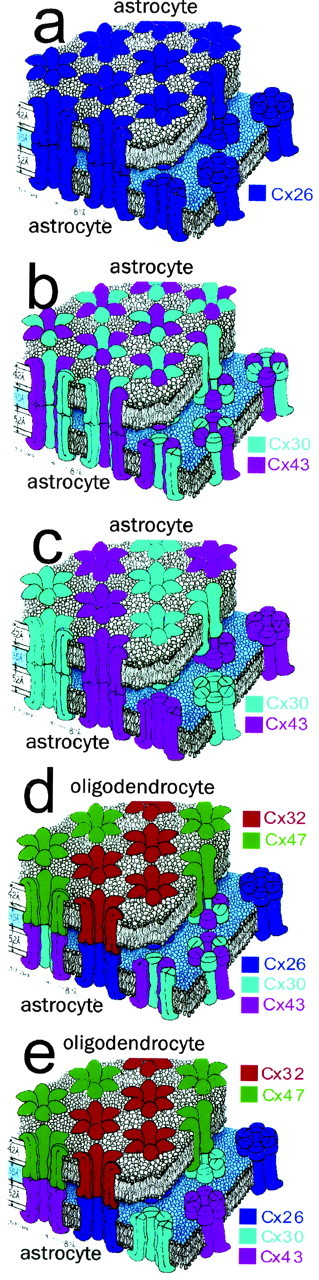
Astrocytes form extensive gap junctions with other astrocytes and with oligodendrocytes. Junctional communication between CNS glia is likely of critical importance because loss of the gap junction channel-forming proteins, connexins Cx32 and Cx47, result in severe demyelination. However, CNS glia express at least six connexins, and the cellular origins and relationships of these proteins have not been determined. We produced a Cx29 reporter mouse in which the connexin coding sequence was replaced with a histological marker, which was used to demonstrate that Cx29, Cx32, and Cx47 are expressed specifically in oligodendrocytes. To determine the relationships between astrocyte and oligodendrocyte connexins, we used double- and triple-immunofluorescence microscopy using semithin sections (<1 microm) of adult mouse spinal cord. Astrocytes form two distinct classes of gap junctions with each other; those composed of Cx26 and those composed of Cx43 and Cx30. In addition, astrocytes establish two classes of intercellular channels with oligodendrocytes, heterotypic Cx26-Cx32 channels and heterotypic Cx30/Cx43-Cx47 channels that may also be heteromeric. In contrast, Cx29 does not colocalize with any of the other five connexins. The data provide the first in vivo demonstration of heterotypic intercellular channels and reveal an unexpected complexity in the composition of glial gap junctions.


Figures

Figure 1.
Figure 1.
Cx29 reporter mouse reveals widespread expression of Cx29. A, Structure of wild-type, targeting vector, and mutant alleles. The Cx29 coding region and ∼800 bp of 3′ untranslated region were replaced with a reporter cassette containing nuclear-localized LacZ. B, Homologous recombination in WT, heterozygote (HET), and KO was verified by PCR. C, Loss of Cx29 in KO spinal cord was verified by Western blot. D, Sagittal section of heterozygote brain at low magnification stained forβ-gal (purple). High levels are observed in all white matter tracts and gray matter regions, as well as the Purkinje cell layer of the cerebellum. E, High-magnification view of boxed region double stained for parvalbumin (green), a marker for Purkinje neurons, andβ-gal (red) reporter. No overlap is evident, suggesting that Cx29 is expressed by Bergmann glia. Scale bar, 10 μm.
Figure 2.
Figure 2.
The majority of oligodendrocytes simultaneously transcribe Cx29, Cx32, and Cx47. A, In heterozygote spinal cord, β-gal (red) was observed in every CC1-positive (green) cell. Note the small soma consisting of large nucleus (red) and scant cytoplasm (green), typical of oligodendrocytes. Cx32-positive (B) and Cx47-positive (C) puncta outline typical β-gal-positive cells and are distributed along proximal process. Scale bars, 10 μm.
Figure 3.
Figure 3.
Oligodendrocyte Cx29 does not associate with other oligodendrocyte connexins. Cross section of WT spinal cord at low magnification triple stained for Cx29, Cx32, and Cx47. Cx29 (blue) is more abundant in the gray matter (GM) than in white matter (WM). Cx32 (red) and Cx47 (green) are colocalized at oligodendrocyte cell bodies in gray matter (box) and white matter (arrow). High-magnification views of the area enclosed by the white dashed box are present at the bottom of the figure. Cx29 (blue) is distributed independently from Cx32 (red) and Cx47 (green), which almost completely overlap (yellow). All nuclei are visualized by DAPI (pseudo-white). Scale bar, 10 μm.
Figure 4.
Figure 4.
There are at least two classes of astrocyte gap junctions, those containing Cx26 and those containing Cx43 and Cx30. A, At low magnification, Cx30 (red) and Cx43 (green) are relatively more abundant in gray matter than in white matter. The boxed area is displayed in the high-magnification panels on the right, which reveal that Cx30 and Cx43 are primarily colocalized (yellow). Because the ratio of Cx30 to Cx43 was variable, not every puncta in the merged image appears yellow. However, careful inspection of the separate channels reveals that the majority of puncta contain both connexins (arrows). B, Alternatively, Cx26 (red) and Cx43 (green) are mostly not colocalized. In addition, Cx43 puncta are more numerous than those containing Cx26. C, As predicted from the above results, Cx26 (green) and Cx30 (red) rarely colocalize. All nuclei are visualized by DAPI (pseudo-white). Scale bars, 10 μm.
Figure 5.
Figure 5.
Oligodendrocyte Cx29 does not associate with any astrocyte connexin. There is no obvious overlap between oligodendrocyte Cx29 (red) and astrocyte Cx26 (blue) or Cx43 (green) at low magnification in any region of the spinal cord. This is confirmed at higher magnification of the boxed area. All nuclei are visualized by DAPI (pseudo-white). GM, Gray matter; WM, white matter. Scale bar, 10 μm.
Figure 6.
Figure 6.
Astrocyte Cx43 preferentially associates with oligodendrocyte Cx47. Triple labeling for Cx32 (red), Cx43 (blue), and Cx47 (green) in adult WT spinal cord demonstrates a high degree of overlap of all three connexins (white puncta) at oligodendrocyte cell bodies and proximal processes in the gray matter. Box A illustrates a typical oligodendrocyte cell body in which all three connexins are colocalized. Box B illustrates a rare oligodendrocyte cell body in which Cx47 and Cx32 do not colocalize. In these circumstances, Cx43 associates only with Cx47 (cyan) and not with Cx32. All nuclei are visualized by DAPI (pseudo-white). Scale bar, 10 μm.
Figure 7.
Figure 7.
Astrocyte Cx30 preferentially associates with oligodendrocyte Cx47. Triple labeling for Cx32 (red), Cx30 (blue), and Cx47 (green) adult WT spinal cord demonstrates a high degree of overlap of all three connexins (white puncta) at oligodendrocyte cell bodies and proximal processes in the gray matter. To determine the oligodendrocyte partner of Cx30, we searched for examples in which oligodendrocyte Cx32 and Cx47 did not overlap (box). Extensive overlap of Cx47 (green) with Cx30 (blue) was observed (cyan), whereas Cx32 (red) was unassociated with Cx30. All nuclei are visualized by DAPI (pseudo-white). Scale bar, 10 μm.
Figure 8.
Figure 8.
Astrocyte Cx26 preferentially associates with oligodendrocyte Cx32. Triple labeling for Cx32 (red), Cx26 (green), and Cx47 (blue) demonstrates a high degree of overlap of all three connexins (white puncta) at gray matter (GM) oligodendrocyte cell bodies (white arrows) and proximal processes. However, in the white matter (WM), oligodendrocyte Cx32 and Cx47 are less often colocalized than in gray matter. In these instances, astrocyte Cx26 (boxed area) is always associated with oligodendrocyte Cx32 (yellow) but not with oligodendrocyte Cx47. All nuclei are visualized by DAPI (pseudo-white). Scale bar, 10 μm.
Figure 9.
Figure 9.
Redistribution of astrocyte connexins at oligodendrocyte cell bodies in Cx32 KO mice. Our data suggest that colocalization of Cx47 and Cx26, observed at oligodendrocyte cell bodies (Fig. 7, low magnification), reflects the heterotypic association of Cx47 with Cx43/Cx30 and of Cx26 with Cx32. If so, then Cx26 should no longer be colocalized with Cx47 at oligodendrocyte cell bodies in the Cx32 KO. Indeed, colocalization (yellow) of Cx26 (red) and Cx47 (green) (column 1, WT) is lost in the Cx32 KO (column 1, Cx32 KO). Also as predicted, Cx43 (red) continues to colocalize (yellow) with Cx47 (green) at oligodendrocyte cell bodies in Cx32 KO (column 2). Unexpectedly, colocalization of Cx30 (red) and Cx47 (green) (column 3) at oligodendrocyte cell bodies is lost in the Cx32 KO, indicating that Cx43 and Cx30 are no longer heteromeric partners (see Results). All nuclei are visualized by DAPI (pseudo-white). Scale bar, 10 μm.
Figure 10.
Figure 10.
Multiple types of gap junctions and intercellular channels formed by astrocytes and oligodendrocytes. Astrocytes form two classes of gap junctions with each other. One class contains only Cx26 (a), whereas a second class contain only Cx30 and Cx43 (b, c). It has not yet been determined whether Cx30 and Cx43 form heteromeric connexons. If so, then the second class of astrocyte junction likely contains a single type of heteromeric, heterotypic intercellular channel (b). If not, then they could contain several types of intercellular channels (c). In contrast, gap junctions between astrocytes and oligodendrocytes are not separated into obligatory classes. However, all astrocyte– oligodendrocyte gap junctions contain heterotypic channels (d, e). Astrocyte Cx26 preferentially associates with oligodendrocyte Cx32, whereas astrocyte Cx30 and Cx43 preferentially associate with oligodendrocyte Cx47. It remains to be determined whether Cx47 interacts with heteromeric connexons containing both Cx30 and Cx43 (d) or whether Cx30 and Cx43 are separated (e). Adapted from Makowski et al. (1977).

References

    1. Altevogt BM, Kleopa KA, Postma FR, Scherer SS, Paul DL (2002) Connexin29 is uniquely distributed within myelinating glial cells of the central and peripheral nervous systems. J Neurosci 22: 6458–6470. - PMC - PubMed
    1. Alvarez-Maubecin V, Garcia-Hernandez F, Williams JT, Van Bockstaele EJ (2000) Functional coupling between neurons and glia. J Neurosci 20: 4091–4098. - PMC - PubMed
    1. Balice-Gordon RJ, Bone LJ, Scherer SS (1998) Functional gap junctions in the Schwann cell myelin sheath. J Cell Biol 142: 1095–1104. - PMC - PubMed
    1. Belliveau DJ, Naus CCG (1994) Cortical type 2 astrocytes are not dye coupled nor do they express the major gap junction genes found in the central nervous system. Glia 12: 24–34. - PubMed
    1. Bergoffen J, Scherer SS, Wang S, Scott MO, Bone LJ, Paul DL, Chen K, Lensch MW, Chance PF, Fischbeck KH (1993) Connexin mutations in X-linked Charcot-Marie-Tooth disease. Science 262: 2039–2042. - PubMed

Publication types

MeSH terms

LinkOut - more resources