

CD4-positive T cell-mediated neuroprotection requires dual compartment antigen presentation
- PMID: 15128847
- PMCID: PMC2665301
- DOI: 10.1523/JNEUROSCI.5276-03.2004
CD4-positive T cell-mediated neuroprotection requires dual compartment antigen presentation
Abstract
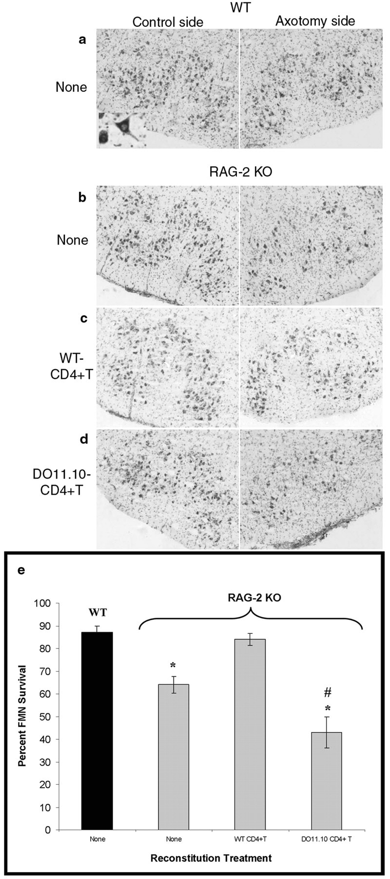
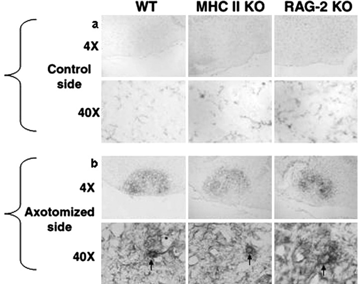
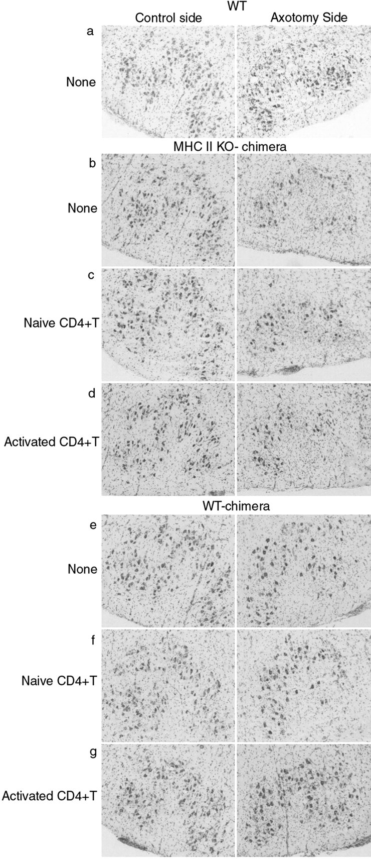
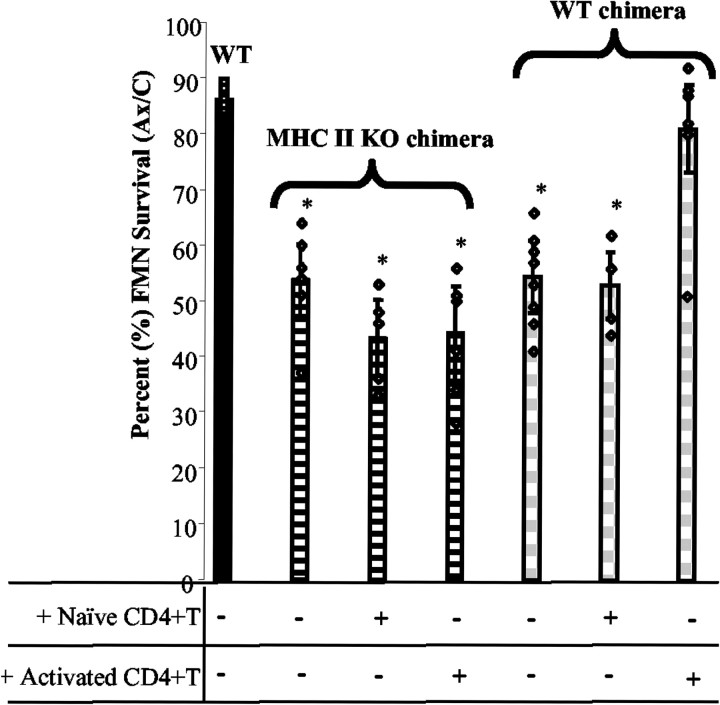
Our laboratory discovered that CD4-positive (CD4+) T cells of the immune system convey transitory neuroprotection to injured mouse facial motoneurons (FMNs) (Serpe et al., 1999, 2000, 2003). A fundamental question in the mechanisms responsible for neuroprotection concerns the identity of the cell(s) that serves as the antigen-presenting cell (APC) to activate the CD4+ T cells. Here, we first establish that CD4+ T cells reactive to non-CNS antigen fail to support FMN survival and, second, demonstrate a two-compartment model of CD4+ T cell activation. Mouse bone marrow (BM) chimeras were developed that discriminate between resident antigen-presenting host cell and BM-derived antigen-presenting donor cell expression of major histocompatibility complex II within central and peripheral compartments, respectively. After facial nerve transection, neither compartment alone is sufficient to result in activated CD4+ T cell-mediated FMN survival. Rather, CD4+ T cell-mediated neuroprotection appears to depend on both resident microglial cells in the central compartment and a BM-derived APC in the peripheral compartment. This is the first in vivo report demonstrating a neuroprotective mechanism requiring APC functions by resident (i.e., parenchymal) microglial cells.
Figures
References
-
- Aloisi F (2001) Immune function of microglia. Glia 36: 165–179. - PubMed
-
- Aloisi F, Ria F, Adorini L (2000) Regulation of T-cell responses by CNS antigen-presenting cells: different roles for microglia and astrocytes. Immunol Today 21: 141–147. - PubMed
-
- Becher B, Prat A, Antel JP (2000) Brain-immune connection: immunoregulatory properties of CNS-resident cells. Glia 29: 293–304. - PubMed
-
- Bretscher P (1992) The two-signal model of lymphocyte activation twenty-one years later. Immunol Today 13: 74–76. - PubMed
-
- Byram SC, Serpe CJ, Pruett SB, Sanders VM, Jones KJ (2003) Natural killer cells do not mediate facial motoneuron survival after facial nerve transection. Brain Behav Immun 17: 417–425. - PubMed
Publication types
MeSH terms
Substances
Grants and funding
LinkOut - more resources
Full Text Sources
Molecular Biology Databases
Research Materials