Memory impairment in aged primates is associated with focal death of cortical neurons and atrophy of subcortical neurons
- PMID: 15128851
- PMCID: PMC6729447
- DOI: 10.1523/JNEUROSCI.4289-03.2004
Memory impairment in aged primates is associated with focal death of cortical neurons and atrophy of subcortical neurons
Abstract
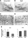
Mechanisms of cognitive decline with aging remain primarily unknown. We determined whether localized cell loss occurred in brain regions associated with age-related cognitive decline in primates. On a task requiring the prefrontal cortex, aged monkeys were impaired in maintaining representations in working memory. Stereological quantification in area 8A, a prefrontal region associated with working memory, demonstrated a significant 32 +/- 11% reduction in the number of Nissl-stained neurons compared with young monkeys. Furthermore, the number of immunolabeled cholinergic neurons projecting to this region of cortex from the nucleus basalis was also reduced by 50 +/- 6%. In contrast, neuronal number was strikingly preserved in an adjoining prefrontal cortical region also associated with working memory, area 46, and in the component of the nucleus basalis projecting to this region. These findings demonstrate extensive but highly localized loss of neocortical neurons in aged, cognitively impaired monkeys that likely contributes to cognitive decline. Cell degeneration, when present, extends transneuronally.
Figures


Similar articles
-
Selective Loss of Thin Spines in Area 7a of the Primate Intraparietal Sulcus Predicts Age-Related Working Memory Impairment.J Neurosci. 2018 Dec 5;38(49):10467-10478. doi: 10.1523/JNEUROSCI.1234-18.2018. Epub 2018 Oct 24. J Neurosci. 2018. PMID: 30355632 Free PMC article.
-
Age-associated neuronal atrophy occurs in the primate brain and is reversible by growth factor gene therapy.Proc Natl Acad Sci U S A. 1999 Sep 14;96(19):10893-8. doi: 10.1073/pnas.96.19.10893. Proc Natl Acad Sci U S A. 1999. PMID: 10485922 Free PMC article.
-
Preservation of prefrontal cortical volume in behaviorally characterized aged macaque monkeys.Exp Neurol. 1999 Nov;160(1):300-10. doi: 10.1006/exnr.1999.7192. Exp Neurol. 1999. PMID: 10630214
-
Life and death of neurons in the aging cerebral cortex.Int Rev Neurobiol. 2007;81:41-57. doi: 10.1016/S0074-7742(06)81004-4. Int Rev Neurobiol. 2007. PMID: 17433917 Review.
-
Neuronal and morphological bases of cognitive decline in aged rhesus monkeys.Age (Dordr). 2012 Oct;34(5):1051-73. doi: 10.1007/s11357-011-9278-5. Epub 2011 Jun 28. Age (Dordr). 2012. PMID: 21710198 Free PMC article. Review.
Cited by
-
Impact of aging brain circuits on cognition.Eur J Neurosci. 2013 Jun;37(12):1903-15. doi: 10.1111/ejn.12183. Eur J Neurosci. 2013. PMID: 23773059 Free PMC article. Review.
-
Recent Insights on the Role of Nuclear Receptors in Alzheimer's Disease: Mechanisms and Therapeutic Application.Int J Mol Sci. 2025 Jan 30;26(3):1207. doi: 10.3390/ijms26031207. Int J Mol Sci. 2025. PMID: 39940973 Free PMC article. Review.
-
Amyloidosis increase is not attenuated by long-term calorie restriction or related to neuron density in the prefrontal cortex of extremely aged rhesus macaques.Geroscience. 2020 Dec;42(6):1733-1749. doi: 10.1007/s11357-020-00259-0. Epub 2020 Sep 2. Geroscience. 2020. PMID: 32876855 Free PMC article.
-
Behavioural phenotyping, learning and memory in young and aged growth hormone-releasing hormone-knockout mice.Endocr Connect. 2018 Aug 1;7(8):924-931. doi: 10.1530/EC-18-0165. Endocr Connect. 2018. PMID: 30300535 Free PMC article.
-
Aging reduces total neuron number in the dorsal component of the rodent prefrontal cortex.J Comp Neurol. 2012 Apr 15;520(6):1318-26. doi: 10.1002/cne.22790. J Comp Neurol. 2012. PMID: 22020730 Free PMC article.
References
-
- Ahmad A, Spear PD (1993) Effects of aging on the size, density, and number of rhesus monkey lateral geniculate neurons. J Comp Neurol 334: 631–643. - PubMed
-
- Baddeley A (1998) Recent developments in working memory. Curr Opin Neurobiol 8: 234–238. - PubMed
-
- Barbas H (2000) Connections underlying the synthesis of cognition, memory, and emotion in primate prefrontal cortices. Brain Res Bull 52: 319–330. - PubMed
-
- Bauer RH, Fuster JM (1976) Delayed-matching and delayed-response deficit from cooling dorsolateral prefrontal cortex in monkeys. J Comp Physiol Psychol 90: 293–302. - PubMed
