. 2004 May 5;24(18):4401-11.
doi: 10.1523/JNEUROSCI.0348-04.2004.

Variant brain-derived neurotrophic factor (BDNF) (Met66) alters the intracellular trafficking and activity-dependent secretion of wild-type BDNF in neurosecretory cells and cortical neurons

Affiliations

Variant brain-derived neurotrophic factor (BDNF) (Met66) alters the intracellular trafficking and activity-dependent secretion of wild-type BDNF in neurosecretory cells and cortical neurons

Zhe-Yu Chen et al. J Neurosci. .

Abstract

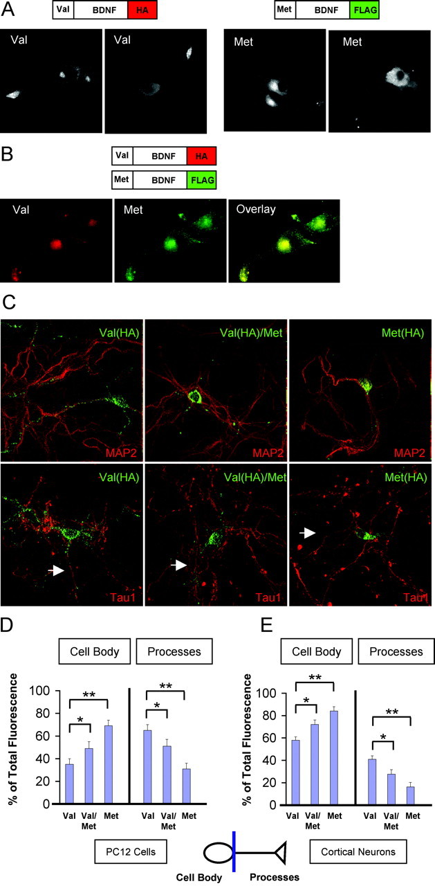
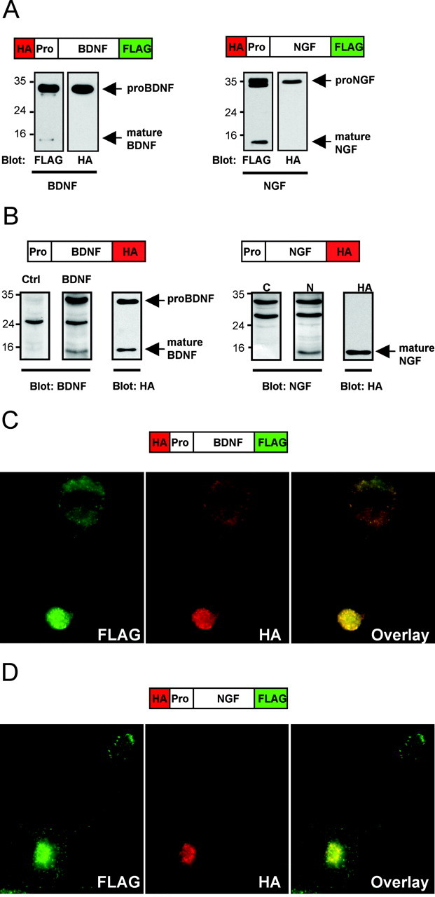
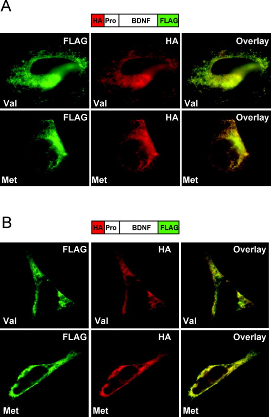
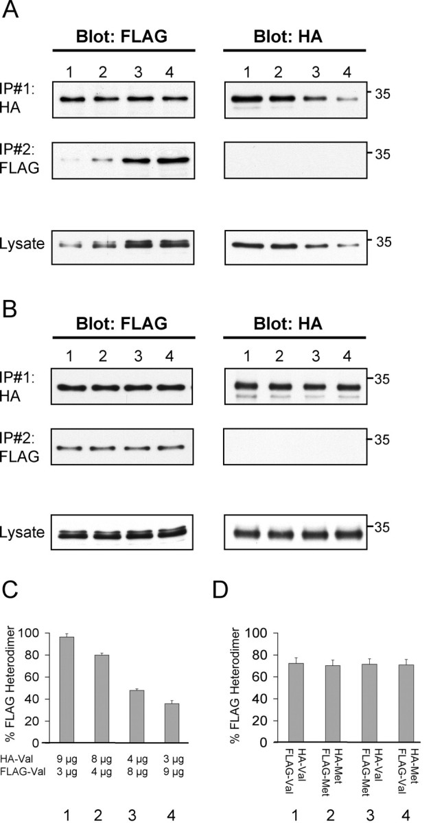
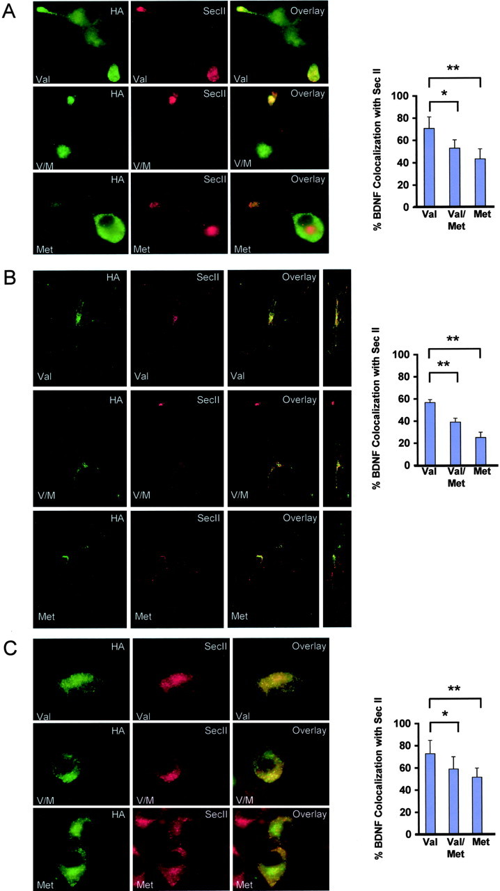
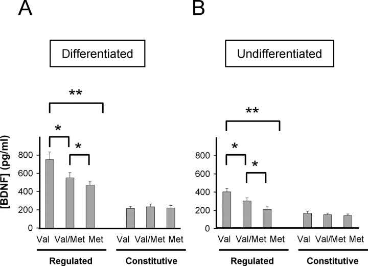
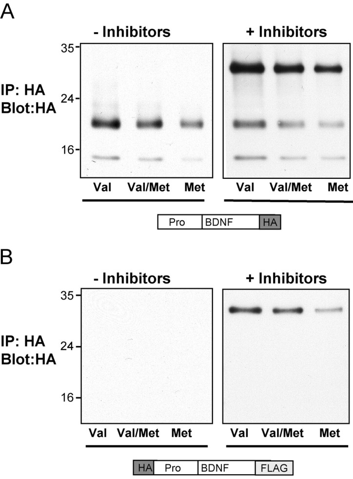
Brain-derived neurotrophic factor (BDNF) plays a critical role in nervous system and cardiovascular development and function. Recently, a common single nucleotide polymorphism in the bdnf gene, resulting in a valine to methionine substitution in the prodomain (BDNF(Met)), has been shown to lead to memory impairment and susceptibility to neuropsychiatric disorders in humans heterozygous for the variant BDNF. When expressed by itself in hippocampal neurons, less BDNF(Met) is secreted in an activity-dependent manner. The nature of the cellular defect when both BDNF(Met) and wild-type BDNF (BDNF(Val)) are present in the same cell is not known. Given that this is the predominant expression profile in humans, we examined the effect of coexpressed BDNF(Met) on BDNF(Val) intracellular trafficking and processing. Our data indicate that abnormal trafficking of BDNF(Met) occurred only in neuronal and neurosecretory cells and that BDNF(Met) could alter the intracellular distribution and activity-dependent secretion of BDNF(Val). We determined that, when coexpressed in the same cell, approximately 70% of the variant BDNF forms BDNF(Val).BDNF(Met) heterodimers, which are inefficiently sorted into secretory granules resulting in a quantitative decreased secretion. Finally, we determined the form of BDNF secreted in an activity-dependent manner and observed no differences in the forms of BDNF(Met) or the BDNF(Val).BDNF(Met) heterodimer compared with BDNF(Val). Together, these findings indicate that components of the regulated secretory machinery interacts specifically with a signal in the BDNF prodomain and that perturbations in BDNF trafficking may lead to selective impairment in CNS function.


Figures

Figure 4.
Figure 4.
Processing and localization of variant and wild-type BDNF. A, Differentiated PC12 cells were transfected with BDNFVal (Val) or BDNFMet (Met) containing a C-terminal HA epitope or C-terminal FLAG epitope tag, respectively. Cells were fixed after 48 hr and permeabilized, and subcellular distribution of neurotrophins was visualized by indirect immunofluorescence microscopy, as described in Materials and Methods, using ant-HA or anti-FLAG antibodies. Representative epifluorescence images are shown. B, Differentiated PC12 cells were transfected with both BDNFVal containing a C-terminal HA epitope and BDNFMet containing a C-terminal FLAG epitope. Cells were fixed after 48 hr and permeabilized, and subcellular distribution of neurotrophins was visualized by indirect immunofluorescence microscopy using epitope antibodies. Representative epifluorescence images are shown. C, Cortical neurons were transfected with BDNFVal (Val) containing a C-terminal HA epitope, BDNFMet (Met) containing a C-terminal FLAG epitope, or both BDNFVal containing a C-terminal HA epitope and BDNFMet containing a C-terminal FLAG epitope (Val/Met). Cells were fixed after 48 hr and permeabilized, and subcellular distribution of neurotrophins was visualized by confocal microscopy, as described in Materials and Methods, using epitope antibodies. In the case of the coexpressed BDNFVal and BDNFMet, staining for the HA epitope was performed. In addition, all neurons were costained with either MAP2 or Tau1 to identify BDNF localization in dendrites and axons (white arrow), respectively. Representative reconstructed images are shown. D, Quantitative analysis of PC12 cell results illustrated in B, as described in Materials and Methods. The proportion of total fluorescence localized to the cell body or processes is presented as a mean ± SEM determined from analysis of four independent experiments. (*p < 0.01; **p < 0.001; Student's t test). E, Quantitative analysis of cortical neuron results illustrated in C, as described in Materials and Methods. The proportion of total fluorescence localized to the cell body or processes is presented as a mean ± SEM determined from analysis of four independent experiments. (*p < 0.01; **p < 0.001; Student's t test).
Figure 1.
Figure 1.
Processing and localization of epitope-tagged neurotrophins. A, Dual N-terminal HA-tagged and C-terminal FLAG-tagged BDNF or NGF was transfected into COS-7 cells, and, after 48 hr, lysates were prepared as described in Materials and Methods and analyzed by immunoblotting with anti-HA and anti-FLAG antibodies. B, Single C-terminal HA-tagged BDNF or NGF was transfected into differentiated PC12 cells. After 48 hr, lysates were prepared as described in Materials and Methods and analyzed by immunoblotting with C-terminal antibodies for BDNF or NGF or with anti-HA antibodies. Dual-epitope-tagged BDNF (C) and NGF (D) were transfected in differentiated PC12 cells. After 48 hr, cells were fixed and permeabilized, and subcellular distribution of neurotrophins was visualized by indirect immunofluorescence microscopy using epitope antibodies. Representative epifluorescence images are shown.
Figure 2.
Figure 2.
Variant BDNF has no defect in localization and secretion in endothelial and vascular smooth muscle cells. A, Localization of dual-epitope-tagged BDNF species in endothelial cells. After transfection, fixation, and permeabilization, subcellular distribution of wild-type (Val) and variant (Met) BDNF was visualized by indirect immunofluorescence microscopy using anti-HA and anti-FLAG antibodies. B, Localization of dual-epitope-tagged BDNFVal in vascular smooth muscle cells. After transfection, fixation, and permeabilization, subcellular distribution of BDNFVal (Val) and variant (Met) BDNF was visualized by indirect immunofluorescence microscopy using anti-HA and anti-FLAG antibodies.
Figure 3.
Figure 3.
Coimmunoprecipitation of BDNFVal and BDNFMet heterodimers. A, To detect different degrees of heterodimerizatiom of two epitope-tagged forms of wild-type BDNF, COS-7 cells were transfected with two wild-type BDNF constructs bearing different C-terminal epitopes (HA or FLAG). The amount of cotransfected FLAG-BDNF and HA-BDNF was varied while maintaining a constant amount of total DNA (12 μg) transfected. Cell lysates were prepared as described in Materials and Methods, and the first immunoprecipitations were performed with HA antibodies (IP#1 indicates HA). A second immunoprecipitation was subsequently performed on the remaining lysates with FLAG antibodies (IP#2 indicates FLAG), and immunoprecipitated samples were analyzed by Western blot. In addition, initial lysates (20 μg of total protein) were analyzed by Western blot for levels of transfected FLAG-BDNF and HA-BDNF. B, To detect different degrees of heterodimerizatiom of epitope-tagged forms of wild-type and variant BDNF, COS-7 cells were transfected with combinations of BDNFVal and BDNFMet constructs bearing different C-terminal epitopes (HA-BDNFVal plus FLAG-BDNFVal, HA-BDNFMet plus FLAG-BDNFMet, HA-BDNFVal plus FLAG-BDNFMet, or FLAG-BDNFVal plus HA-BDNFMet). A constant amount (6 μg) of each epitope-tagged BDNF species was transfected to maintain a constant amount of total DNA (12 μg) transfected. Cell lysates were prepared as described in Materials and Methods, and sequential immunoprecipitations were performed (IP#1 indicates HA; IP#2 indicates FLAG), as described in A, and analyzed by Western blot. In addition, initial lysates (20 μg of total protein) were analyzed by Western blot for levels of transfected BDNFVal and BDNFMet. C, Quantitation of Western blots in A as a proportion of FLAG-BDNF immunoreactivity found in IP#1 over the total FLAG immunoreactivity obtained from both immunoprecipitation steps (IP#1 and IP#2, Blot: FLAG). Mean ± SEM proportions were determined from analysis of three independent experiments. D, Quantitation of Western blots in B as a proportion of FLAG-BDNF immunoreactivity found in IP#1 over the total FLAG immunoreactivity obtained from both immunoprecipitation steps (IP#1 and IP#2, Blot: FLAG). Mean ± SEM proportions were determined from analysis of three independent experiments.
Figure 5.
Figure 5.
Subcellular colocalization of variant and wild-type BDNF with organelle markers. Differentiated PC12 cells (A), cortical neurons (B), and undifferentiated PC12 cells (C) were transfected with C-terminal HA-tagged variant (Met), C-terminal HA-tagged wild-type BDNF (Val), or both C-terminal FLAG-tagged variant and HA-tagged wild-type BDNF (V/M). Cells were fixed after 48 hr and permeabilized. Colocalization of BDNF and the secretory granule marker SecII was visualized by costaining with anti-HA and anti-SecII antibodies. Confocal three-dimensional reconstruction of serial z planes were obtained, and alternate rotation angle is also shown for each representative cell. Quantitative analysis of results illustrated in A–C was conducted by scoring ∼25 cells for each BDNF condition. The proportion of colocalization between BDNF and SecII is presented as a mean ± SEM determined from analysis of four independent experiments (*p < 0.01; **p < 0.001; Student's t test).
Figure 6.
Figure 6.
Regulated and constitutive secretion of BDNFVal and BDNFMet. Differentiated (A) and undifferentiated (B) PC12 cells were transfected with BDNFVal (Val), BDNFMet (Met), or both BDNF species (Val/Met). After 48 hr, media was collected under depolarization and constitutive secretion conditions, as described in Materials and Methods, and analyzed by ELISA. Results are presented as a mean ± SEM determined from analysis of three independent experiments (*p < 0.01; **p < 0.001; Student's t test).
Figure 7.
Figure 7.
Identification of secreted forms of BDNFMet. Differentiated PC12 cells were transfected with BDNFVal (Val), BDNFMet (Met), or both BDNFVal and BDNFMet (Val/Met). The BDNF constructs contained either a single C-terminal HA epitope tag (A) or dual N-terminal HA and FLAG C-terminal tags (B). After 48 hr, media was collected under depolarization conditions, as described in Materials and Methods, in the absence or presence of protease inhibitors. BDNF in the media was immunoprecipitated with polyclonal anti-HA antibodies and analyzed by immunoblotting with monoclonal anti-HA antibodies. Representative blots from three independent experiments are shown.

References

    1. Beattie MS, Harrington AW, Lee R, Kim JY, Boyce SL, Longo FM, Bresnahan JC, Hempstead BL, Yoon SO (2002) ProNGF induces p75-mediated death of oligodendrocytes following spinal cord injury. Neuron 36: 375–386. - PMC - PubMed
    1. Chao MV (2003) Neurotrophins and their receptors: a convergence point for many signalling pathways. Nat Rev Neurosci 4: 299–309. - PubMed
    1. Donovan M, Miranda R, Kraemer R, McCaffrey T, Tessarollo L, Mahadeo D, Sharif S, Kaplan D, Tsoulfas P, Parada L, Hempstead B (1995) Neurotrophin and neurotrophin receptors in vascular smooth muscle cells. Regulation of expression in response to injury. Am J Pathol 147: 309–324. - PMC - PubMed
    1. Donovan M, Lin M, Wiegn P, Ringstedt T, Kraemer R, Hahn R, Wang S, Ibanez C, Rafii S, Hempstead B (2000) Brain derived neurotrophic factor is an endothelial cell survival factor required for intramyocardial vessel stabilization. Development 127: 4531–4540. - PubMed
    1. Egan M, Kojima M, Callicott J, Goldberg T, Kolachana B, Bertolino A, Zaitsev E, Gold B, Goldman D, Dean M, Lu B, Weinberger D (2003) The BDNF val66met polymorphism affects activity-dependent secretion of BDNF and human memory and hippocampal function. Cell 112: 257–269. - PubMed

Publication types

MeSH terms

Substances