Biliary atresia is associated with CD4+ Th1 cell-mediated portal tract inflammation
- PMID: 15128911
- PMCID: PMC1948976
- DOI: 10.1203/01.PDR.0000130480.51066.FB
Biliary atresia is associated with CD4+ Th1 cell-mediated portal tract inflammation
Abstract
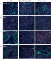
A proposed mechanism in the pathogenesis of biliary atresia involves an initial virus-induced, progressive T cell-mediated inflammatory obliteration of bile ducts. The aim of this study was to characterize the inflammatory environment present within the liver of infants with biliary atresia to gain insight into the role of a primary immune-mediated process versus a nonspecific secondary response to biliary obstruction. Frozen liver tissue obtained from patients with biliary atresia, neonatal giant cell hepatitis, total parenteral nutrition (TPN)-related cholestasis, choledochal cysts, and normal control subjects was used for fluorescent immunohistochemistry studies of cellular infiltrates, cytokine mRNA expression, and in situ hybridization for localization of cytokine-producing cells. Immunohistochemistry revealed increases in CD8(+) and CD4(+) T cells and Kupffer cells (CD68(+)) in the portal tracts of biliary atresia. Reverse transcription-PCR analysis of biliary atresia tissue showed a Th1-type cytokine profile with expression of IL-2, interferon-gamma, tumor necrosis factor-alpha, and IL-12. This profile was not seen in normal, neonatal hepatitis or choledochal cyst livers but was present in TPN-related cholestasis. In situ hybridization revealed that the Th1 cytokine-producing cells were located in the portal tracts in biliary atresia and in the parenchyma of TPN-related cholestasis. A distinctive portal tract inflammatory environment is present in biliary atresia, involving CD4(+) Th1 cell-mediated immunity. The absence of similar inflammation in other pediatric cholestatic conditions suggests that the portal tract inflammation in biliary atresia is not a secondary response to cholestasis but rather indicates a specific immune response involved in the pathogenesis of biliary atresia.
Figures


Comment in
-
Biliary atresia and Th1 function: linking lymphocytes and bile ducts: commentary on the article by Mack et al. on page 79.Pediatr Res. 2004 Jul;56(1):9-10. doi: 10.1203/01.PDR.0000129655.02381.F0. Epub 2004 May 5. Pediatr Res. 2004. PMID: 15128925 Review. No abstract available.
Similar articles
-
Immune-mediated liver injury: prognostic value of CD4+, CD8+, and CD68+ in infants with extrahepatic biliary atresia.J Pediatr Surg. 2005 Aug;40(8):1252-7. doi: 10.1016/j.jpedsurg.2005.05.007. J Pediatr Surg. 2005. PMID: 16080928
-
Elevated expression of IL2 is associated with increased infiltration of CD8+ T cells in biliary atresia.J Pediatr Surg. 2006 Feb;41(2):300-5. doi: 10.1016/j.jpedsurg.2005.11.044. J Pediatr Surg. 2006. PMID: 16481239
-
Oligoclonal expansions of CD4+ and CD8+ T-cells in the target organ of patients with biliary atresia.Gastroenterology. 2007 Jul;133(1):278-87. doi: 10.1053/j.gastro.2007.04.032. Epub 2007 Apr 20. Gastroenterology. 2007. PMID: 17631149 Free PMC article.
-
Biliary atresia.Semin Immunopathol. 2009 Sep;31(3):371-81. doi: 10.1007/s00281-009-0171-6. Epub 2009 Jun 17. Semin Immunopathol. 2009. PMID: 19533128 Review.
-
Recent progress in the etiopathogenesis of pediatric biliary disease, particularly Caroli's disease with congenital hepatic fibrosis and biliary atresia.Histol Histopathol. 2010 Feb;25(2):223-35. doi: 10.14670/HH-25.223. Histol Histopathol. 2010. PMID: 20017109 Review.
Cited by
-
What Causes Biliary Atresia? Unique Aspects of the Neonatal Immune System Provide Clues to Disease Pathogenesis.Cell Mol Gastroenterol Hepatol. 2015 May 1;1(3):267-274. doi: 10.1016/j.jcmgh.2015.04.001. Cell Mol Gastroenterol Hepatol. 2015. PMID: 26090510 Free PMC article.
-
Polymorphisms of the ICAM-1 gene are associated with biliary atresia.Dig Dis Sci. 2008 Jul;53(7):2000-4. doi: 10.1007/s10620-007-9914-1. Epub 2008 Apr 10. Dig Dis Sci. 2008. PMID: 18401716
-
Aetiology of biliary atresia: what is actually known?Orphanet J Rare Dis. 2013 Aug 29;8:128. doi: 10.1186/1750-1172-8-128. Orphanet J Rare Dis. 2013. PMID: 23987231 Free PMC article. Review.
-
Adjuvant therapy in biliary atresia: hopelessly optimistic or potential for change?Pediatr Surg Int. 2017 Dec;33(12):1263-1273. doi: 10.1007/s00383-017-4157-5. Epub 2017 Sep 22. Pediatr Surg Int. 2017. PMID: 28940004 Review.
-
Exome-wide assessment of isolated biliary atresia: A report from the National Birth Defects Prevention Study using child-parent trios and a case-control design to identify novel rare variants.Am J Med Genet A. 2023 Jun;191(6):1546-1556. doi: 10.1002/ajmg.a.63185. Epub 2023 Mar 21. Am J Med Genet A. 2023. PMID: 36942736 Free PMC article.
References
-
- Stein JE, Vacanti JP. Biliary atresia and other disorders of the extrahepatic biliary tree. In: Suchy FJ, editor. Liver Disease in Children. Mosby; London: 1994. pp. 426–442.
-
- Tan CEL, Driver M, Howard ER, Moscoso GJ. Extrahepatic biliary atresia: a first trimester event? Clues from light microscopy and immunohistochemistry. J Pediatr Surg. 1994;29:808–814. - PubMed
-
- Sokol RJ, Mack CL. Etiopathogenesis of biliary atresia. Semin Liver Dis. 2001;21:517–524. - PubMed
-
- Schreiber RA, Kleinman RE. Genetics, immunology and biliary atresia: an opening or a diversion? J Pediatr Gastroenterol Nutr. 1993;16:111–113. - PubMed
Publication types
MeSH terms
Grants and funding
LinkOut - more resources
Full Text Sources
Research Materials

