

Effects of perfusion on radiofrequency ablation in swine kidneys
- PMID: 15128994
- PMCID: PMC2408952
- DOI: 10.1148/radiol.2312021248
Effects of perfusion on radiofrequency ablation in swine kidneys
Abstract
Purpose: To evaluate the effect of vascular occlusion on the size of radiofrequency (RF) ablation lesions and to evaluate embolization as an occlusion method.
Materials and methods: The kidneys of six swine were surgically exposed. Fifteen RF ablation lesions were created in nine kidneys by using a 2-cm-tip single-needle ablation probe in varying conditions: Seven lesions were created with normal blood flow and eight were created with blood flow obstructed by means of vascular clamping (n = 5) or renal artery embolization (n = 3). The temperature, applied voltage, current, and impedance were recorded during RF ablation. Tissue-cooling curves acquired for 2 minutes immediately after the ablation were compared by using regression analysis. Lesions were bisected, and their maximum diameters were measured and compared by using analysis of variance.
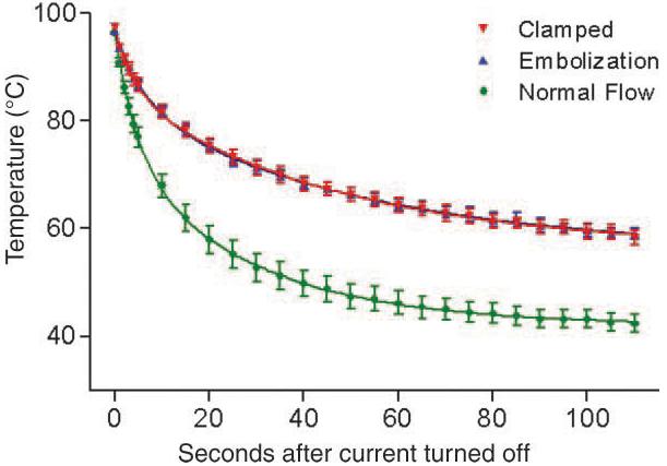
Results: The mean diameter of ablation lesions created when blood flow was obstructed was 60% greater than that of lesions created when blood flow was normal (1.38 cm +/- 0.05 [standard error of mean] vs 0.86 cm +/- 0.07, P <.001). The two methods of flow obstruction yielded lesions of similar mean sizes: 1.40 cm +/- 0.06 with vascular clamping and 1.33 cm +/- 0.07 with embolization. The temperature at the probe tip when lesions were ablated with normal blood flow decreased more rapidly than did the temperature when lesions were ablated after flow obstruction (P <.001), but no significant differences in tissue-cooling curves between the two flow obstruction methods were observed.
Conclusion: Obstruction of renal blood flow before and during RF ablation resulted in larger thermal lesions with potentially less variation in size compared with the lesions created with normal nonobstructed blood flow. Selective arterial embolization of the kidney vessels may be a useful adjunct to RF ablation of kidney tumors.
Figures


Comment in
-
Vascular occlusion: can we push radiofrequency ablation into new size frontiers?Radiology. 2004 May;231(2):291-2. doi: 10.1148/radiol.2312031861. Radiology. 2004. PMID: 15128977 No abstract available.
References
-
- Landis SH, Murray T, Bolden S, Wingo PA. Cancer statistics, 1999. CA Cancer J Clin. 1999;49:8–31. - PubMed
-
- Frank W, Guinan P, Stuhldreher D, Saffrin R, Ray P, Rubenstein M. Renal cell carcinoma: the size variable. J Surg Oncol. 1993;54:163–166. - PubMed
-
- Walther MM, Choyke PL, Glenn G, et al. Renal cancer in families with hereditary renal cancer: prospective analysis of a tumor size threshold for renal parenchymal sparing surgery. J Urol. 1999;161:1475–1479. - PubMed
-
- Smith SJ, Bosniak MA, Megibow AJ, Hulnick DH, Horii SC, Raghavendra BN. Renal cell carcinoma: earlier discovery and increased detection. Radiology. 1989;170:699–703. - PubMed
-
- Bosniak MA, Rofsky NM. Problems in the detection and characterization of small renal masses. Radiology. 1996;200:286–287. - PubMed
Publication types
MeSH terms
Grants and funding
LinkOut - more resources
Full Text Sources
Other Literature Sources
