. 2004 Sep;287(3):G734-41.
doi: 10.1152/ajpgi.00300.2003. Epub 2004 May 6.

Limited role for CXC chemokines in the pathogenesis of alpha-naphthylisothiocyanate-induced liver injury

Affiliations

Limited role for CXC chemokines in the pathogenesis of alpha-naphthylisothiocyanate-induced liver injury

Junquan Xu et al. Am J Physiol Gastrointest Liver Physiol. 2004 Sep.

Abstract

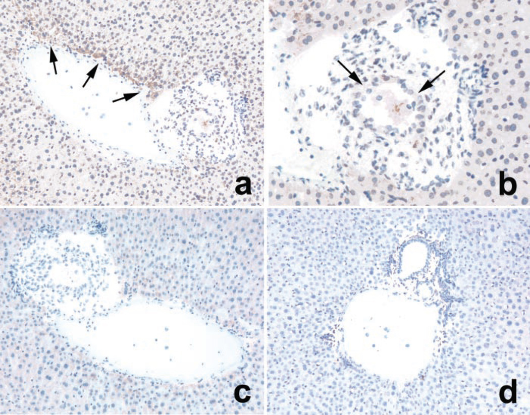
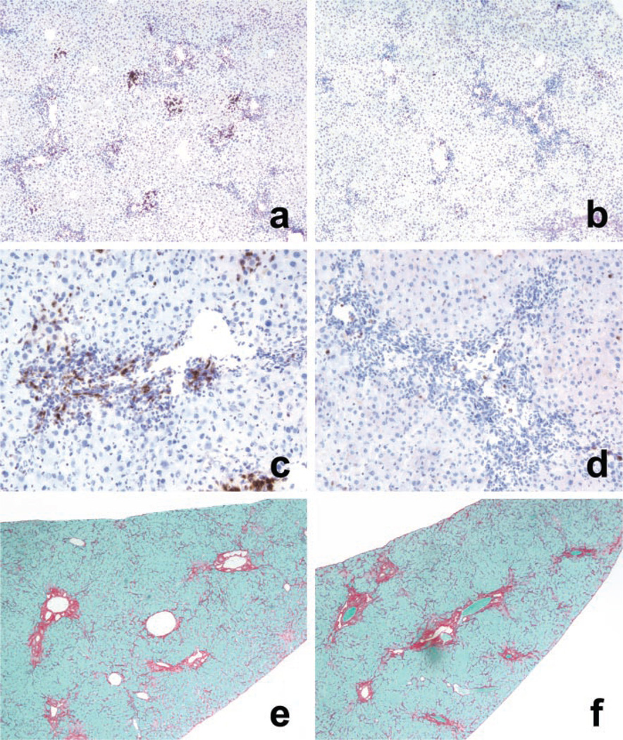
Alpha-naphthylisothiocyanate (ANIT) is a hepatotoxin that causes severe neutrophilic inflammation around portal tracts and bile ducts. The chemotactic signals that provoke this inflammatory response are unknown. In this study, we addressed the possibility that ANIT upregulates CXC chemokines in the liver and that these compounds mediate hepatic inflammation and tissue injury after ANIT treatment. Mice treated with a single dose of ANIT (50 mg/kg) exhibited rapid hepatic induction of the CXC chemokine macrophage inflammatory protein-2 (MIP-2). MIP-2 derived primarily from hepatocytes, with no apparent contribution by biliary cells. In ANIT-treated mice, the induction of MIP-2 coincided with an influx of neutrophils to portal zones; this hepatic neutrophil recruitment was suppressed by 50% in mice that lack the receptor for MIP-2 (CXCR2(-/-)). Interestingly, despite their markedly reduced degree of hepatic inflammation, CXCR2(-/-) mice displayed just as much hepatocellular injury and cholestasis after ANIT treatment as wild-type mice. Moreover, after long-term exposure, ANIT CXCR2(-/-) mice developed liver fibrosis that was indistinguishable from that in wild-type mice. In summary, our data show that CXC chemokines are responsible for some of the hepatic inflammation that occurs in response to ANIT but that these compounds are not essential to the pathogenesis of either acute or chronic ANIT hepatotoxicity.


Figures

Fig. 1
Fig. 1
Induction of macrophage inflammatory protein-2 (MIP-2) in mouse liver after α-naphthylisothiocyanate (ANIT) treatment. Histogram shows the amount of MIP-2 in liver homogenates from wild-type (WT) and CXCR2−/− [knockout (KO)] mice at various intervals after a single dose of ANIT (50 mg/kg). Chemokine levels rose abruptly at 12 h and declined toward normal at 48 h. Values represent means ± SE for n ≥ 4. *P < 0.05 vs. 6-h value of similar genotype. #P < 0.05 vs. 48-h WT mice.
Fig. 2
Fig. 2
Localization of MIP-2 in mouse liver after ANIT treatment. MIP-2 was identified in mouse liver sections by immunohistochemistry (see materials and methods). Twenty-four hours after ANIT, MIP-2 was visible in periportal hepatocytes (a; arrows) but not in biliary epithelial cells (b; arrows). Control sections of ANIT-treated liver, which were incubated with nonimmune IgG in place of anti-MIP-2, showed no staining (c). The livers of control mice that received vehicle instead of ANIT displayed no MIP-2 at 24 h (d).
Fig. 3
Fig. 3
Hepatic neutrophil counts in WT and KO mice after acute ANIT treatment. Histogram illustrates the number of neutrophils present in the livers of WT and KO mice at various intervals after a single dose of ANIT (50 mg/kg). Neutrophils were quantitated by direct counting of tissue sections stained with anti-neutrophil antibody (Ly-6G; see materials and methods). Values represent means ± SE for n ≥ 4. *P < 0.05 vs. 6-h value of similar genotype. #P < 0.05 vs. WT mice at the same interval.
Fig. 4
Fig. 4
Liver histology in WT and KO mice after acute ANIT treatment. Mice received a single dose of ANIT (50 mg/kg) by gavage and were killed 12 h later, the time point at which neutrophils were most numerous. WT mice are represented in a, c, and e; KO mice are represented in b, d, and f. WT mice show robust periductular inflammation in response to ANIT (a; arrowheads). The neutrophilic component of the inflammatory infiltrate is highlighted by Ly-6G immunohistochemistry (c, e). Compared with WT mice, KO mice display less overall biliary ductular inflammation after ANIT treatment (b; arrowheads) as well as a lesser degree of neutrophil infiltration (d, f).
Fig. 5
Fig. 5
Biochemical markers of liver injury in WT and KO mice after acute ANIT treatment. Histograms show serum alanine aminotransferase (ALT), alkaline phosphatase, and total bilirubin in WT and KO mice at various intervals after a single dose of ANIT (50 mg/kg). Values represent means ± SE for n ≥ 4. *P < 0.05 vs. 6-h value of similar genotype.
Fig. 6
Fig. 6
Liver histology in WT and KO mice after chronic ANIT treatment. Photomicrographs illustrate neutrophilic inflammation (a–d) and fibrosis (e, f) in mouse liver after 8 wk of ANIT feeding. a, c, And e are from WT mice; b, d, and f are from KO. Neutrophils are much more prominent in the livers of WT mice than KO mice. WT livers contain clusters of neutrophils scattered throughout the hepatic parenchyma (a). High-power magnification shows that these cells are localized in expanded portal tracts (c). In the livers of KO mice, neutrophils are scarce (b, d). Despite the difference in hepatic inflammation, hepatic fibrosis is similar in both WT and knockout mice (e, f).

References

    1. Baggiolini M. Chemokines and leukocyte traffic. Nature. 1998;392:565–568. - PubMed
    1. Bailie MB, Dahm LJ, Peters-Golden M, Harris RR, Carter GW, Roth RA. Leukotrienes and α-naphthylisothiocyanate-induced liver injury. Toxicology. 1995;100:139–149. - PubMed
    1. Bailie MB, Pearson JM, Fink GD, Roth RA. Platelet activating factor receptor blockade alone or in combination with leukotriene synthesis inhibition does not ameliorate α-naphthylisothiocyanate-induced hepatotoxicity. Toxicol Lett. 1996;84:89–95. - PubMed
    1. Bautista AP. Impact of alcohol on the ability of Kupffer cells to produce chemokines and its role in alcoholic liver disease. J Gastroenterol Hepatol. 2000;15:349–356. - PubMed
    1. Bautista AP. Chronic alcohol intoxication primes Kupffer cells and endothelial cells for enhanced CC-chemokine production and concomitantly suppresses phagocytosis and chemotaxis. Front Biosci. 2002;7:117–125. - PubMed

Publication types

MeSH terms