

Specific detection and identification of herpes B virus by a PCR-microplate hybridization assay
- PMID: 15131142
- PMCID: PMC404616
- DOI: 10.1128/JCM.42.5.1869-1874.2004
Specific detection and identification of herpes B virus by a PCR-microplate hybridization assay
Abstract
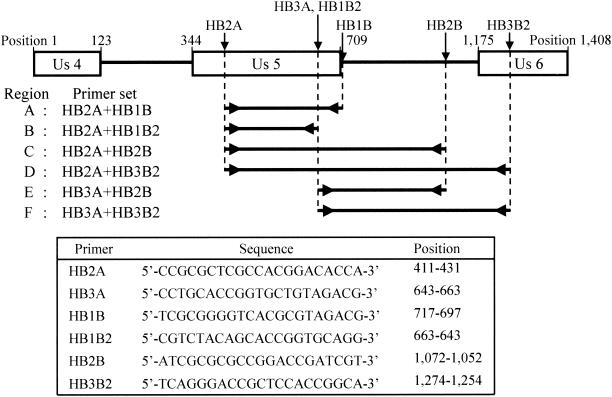
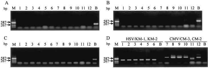
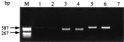
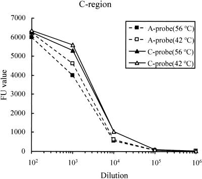
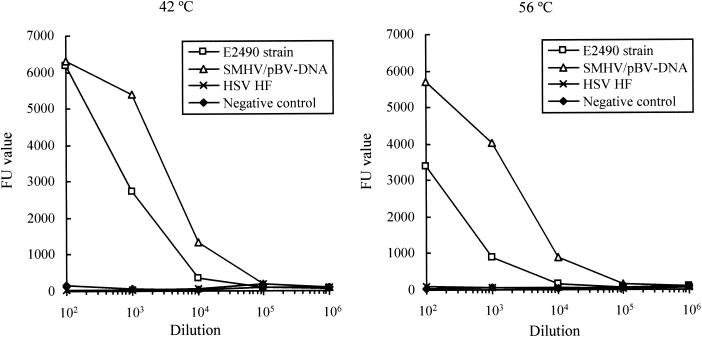
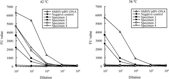
Herpes B virus DNA was specifically amplified by PCR, targeting the regions that did not cross-react with herpes simplex virus (HSV). The amplified products, which were shown to be highly genetic polymorphisms among herpes B virus isolates, were identified by microplate hybridization with probes generated by PCR. The products immobilized in microplate wells were hybridized with the biotin-labeled probes derived from the SMHV strain of herpes B virus. The amplified products derived from the SMHV and E2490 strains of herpes B virus were identified by microplate hybridization. PCR products amplified from the trigeminal ganglia of seropositive cynomolgus macaques were identified as herpes B virus DNA. The utility of the PCR-microplate hybridization assay for genetic detection and identification of the polymorphic region of herpes B virus was determined.
Figures
Similar articles
-
Prevalence of herpes B virus genome in the trigeminal ganglia of seropositive cynomolgus macaques.Lab Anim. 2008 Jan;42(1):99-103. doi: 10.1258/la.2007.006031. Lab Anim. 2008. PMID: 18348771
-
Polymerase chain reaction for detection of herpesvirus simiae (B virus) in clinical specimens.Arch Virol. 1993;131(1-2):89-9. doi: 10.1007/BF01379082. Arch Virol. 1993. PMID: 8392323
-
Rapid detection of B virus (herpesvirus simiae) DNA by polymerase chain reaction.J Infect Dis. 1993 Sep;168(3):747-50. doi: 10.1093/infdis/168.3.747. J Infect Dis. 1993. PMID: 8394866
-
Detection of herpes simplex virus DNA in cerebrospinal fluid samples using the polymerase chain reaction and microplate hybridization.J Virol Methods. 1996 May;59(1-2):1-11. doi: 10.1016/0166-0934(95)01991-x. J Virol Methods. 1996. PMID: 8793825
-
Towards a comprehensive view of the herpes B virus.Front Immunol. 2023 Nov 16;14:1281384. doi: 10.3389/fimmu.2023.1281384. eCollection 2023. Front Immunol. 2023. PMID: 38035092 Free PMC article. Review.
Cited by
-
Pathologic characteristics of infectious diseases in macaque monkeys used in biomedical and toxicologic studies.J Toxicol Pathol. 2023 Apr;36(2):95-122. doi: 10.1293/tox.2022-0089. Epub 2023 Feb 13. J Toxicol Pathol. 2023. PMID: 37101957 Free PMC article. Review.
-
Herpes B virus gD interaction with its human receptor--an in silico analysis approach.Theor Biol Med Model. 2014 Jun 6;11:27. doi: 10.1186/1742-4682-11-27. Theor Biol Med Model. 2014. PMID: 24902525 Free PMC article.
-
Cytomegalovirus distribution and evolution in hominines.Virus Evol. 2019 Aug 1;5(2):vez015. doi: 10.1093/ve/vez015. eCollection 2019 Jul. Virus Evol. 2019. PMID: 31384482 Free PMC article.
-
Evolution and Genetic Diversity of Primate Cytomegaloviruses.Microorganisms. 2020 Apr 25;8(5):624. doi: 10.3390/microorganisms8050624. Microorganisms. 2020. PMID: 32344906 Free PMC article. Review.
-
Specific pathogen free macaque colonies: a review of principles and recent advances for viral testing and colony management.J Med Primatol. 2016 Apr;45(2):55-78. doi: 10.1111/jmp.12209. Epub 2016 Mar 1. J Med Primatol. 2016. PMID: 26932456 Free PMC article. Review.
References
-
- Bennett, A. M., L. Harrington, and D. C. Kelly. 1992. Nucleotide sequence analysis of genes encoding glycoproteins D and J in simian herpes B virus. J. Gen. Virol. 73:2963-2967. - PubMed
-
- Boulter, E. A. 1975. The isolation of monkey B virus (Herpesvirus simiae) from the trigeminal ganglia of a healthy seropositive rhesus monkey. J. Biol. Stand. 3:279-280. - PubMed
-
- Cohen, J. I., D. S. Davenport, J. A. Stewart, S. Deitchman, J. K. Hilliard, L. E. Chapman, et al. 2002. Recommendations for prevention of and therapy for exposure to B virus (Cercopithecine Herpesvirus 1). Clin. Infect. Dis. 35:1191-1203. - PubMed
-
- Eberle, R., D. Black, and J. K. Hilliard. 1989. Relatedness of glycoproteins expressed on the surface of simian herpes-virus virions and infected cells to specific HSV glycoproteins. Arch. Virol. 109:233-252. - PubMed
Publication types
MeSH terms
Substances
LinkOut - more resources
Full Text Sources
