

Comparative Study
doi: 10.1128/JCM.42.5.1982-1985.2004.
Amplification of coccidioidal DNA in clinical specimens by PCR
Affiliations
- PMID: 15131158
- PMCID: PMC404645
- DOI: 10.1128/JCM.42.5.1982-1985.2004
Item in Clipboard
Comparative Study
Amplification of coccidioidal DNA in clinical specimens by PCR
J Clin Microbiol.
2004 May.
Abstract
Coccidioides DNA was amplified from serum by a PCR using coccidioid-specific primers. A 239-bp product was visualized when 10 fg of exogenous coccidioidal DNA was subjected to amplification. This product was demonstrated in some human and mouse sera prior to the detection of coccidioidal antibodies.
Figures
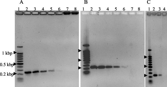
Demonstration of coccidioidal PCR products after agarose gel electrophoresis. A 100-bp DNA ladder was used to estimate the product sizes (lane 1). (A and B) Coccidioidal genomic DNAs were added to PCRs in the following quantities to determine the sensitivity of detetion. Lane 2, 100 pg; lane 3, 10 pg; lane 4, 1 pg; lane 5, 100 fg; lane 6, 10 fg; lane 7, 1 fg; lane 8, no DNA added. Gels were stained with ethidium bromide (A) or Vistra Green (B). (C) Recovery of coccidioidal DNA added to serum. Lane 2, coccidioidal DNA added to normal human serum after the isolation procedure and then amplified by PCR; lane 3, coccidioidal genomic DNA added to normal human serum prior to isolation and PCR amplification; lane 4, negative (no template) control reaction. The gel was stained with ethidium bromide.
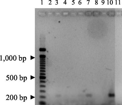
PCR amplification of coccidioidal DNAs from clinical specimens. Products were separated by electrophoresis through a 1.5% agarose gel and stained with ethidium bromide. Lanes 2, 3, 4, 6, 7, 8, and 9, sera that were positive for coccidioidal antibodies; lane 5, seronegative sample; lane 10, 100 fg of coccidioidal DNA as a positive control; lane 11, negative (no template) control.
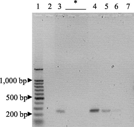
PCR amplification of coccidioidal DNAs from mouse sera. DNAs present in sera collected from mice after infection were amplified, separated by electrophoresis, and stained with Vistra Green. Amplified samples collected 3 days after infection are shown. Lane 2, uninfected mice; lane 3, mice infected with 500 arthroconidia. While samples collected after 3 days were also subjected to amplification and electrophoresis, no products were visible, and representative lanes are indicated by an asterisk. Lane 1, 100-bp DNA ladder; lane 4, 10-pg PCR control; lane 5, 1-pg PCR control; lane 6, 100-fg PCR control; lane 7, negative (no template) control.
Comment in
-
Amplification of coccidioidal DNA in clinical specimens by PCR.J Clin Microbiol. 2005 Mar;43(3):1492; author reply 1492-3. doi: 10.1128/JCM.43.3.1492-1493.2005. J Clin Microbiol. 2005. PMID: 15750145 Free PMC article. No abstract available.
References
-
- Clark, K. A., and D. McAllister. 1996. Direct detection of Coccidioides immitis in clinical specimens using target amplification, p. 129-136. In H. E. Einstein and A. Cantanzaro (ed.), Coccidioidomycosis. Proceedings of the 5th International Conference. National Foundation for Infectious Diseases, Bethesda, Md.
-
- Fisher, M. C., G. L. Koenig, T. J. White, and J. W. Taylor. 2002. Molecular and phenotypic description of Coccidioides posadasii sp. nov., previously recognized as the non-California population of Coccidioides immitis. Mycologia 94:73-84. - PubMed
Publication types
MeSH terms
Substances
LinkOut - more resources
Full Text Sources
Other Literature Sources
